Excipients influence neural responses: an intrathecal citrate animal study
Intrathecal citrate exposure accompanies dose-related sensorimotor abnormalities in mice and rabbits, with early reductions and later increases in mouse cortical activity markers and calcium signals.

Excipients help formulations dissolve, buffer and remain stable. What happens when an excipient enters the cerebrospinal-fluid environment? A Pharmaceutics study examines intrathecal citric acid/sodium citrate, or SC, in mice and rabbits. Higher exposure and repeated administration accompany greater sensorimotor abnormalities, while cortical signals first weaken and later strengthen. A formulation component becomes the focus of investigation.
Repeated intrathecal SC exposure reduces mouse movement and grid-test performance and delays heat responses. PrL signals decrease early and increase later. Higher rabbit doses produce transient abnormalities without evident local structural damage in examined tissue.
Contents · 6 sections
From buffering component to the CSF environment
Intrathecal delivery brings formulations close to the CNS. Different citrate exposures are compared with saline, with repeated administration in mice. Formulation pH and osmolarity are kept near physiological ranges to examine functional effects of exposure.
Sensory tests, movement measures, regional activity and tissue observations address different aspects of the response. Together they describe how a formulation component’s functional effects vary across time and outcomes.
Hindlimb performance changes alongside delayed heat responses
After two SC exposures, higher-dose mice travel less and more slowly in an open field. Several dose groups fall sooner in the grid test, indicating poorer maintenance of grip and posture. The most marked locomotor reduction occurs at higher exposure.
Hot-plate paw-lifting latency increases and thermal-response counts decrease. These outcomes occur alongside motor abnormalities and describe altered sensorimotor function. A slower withdrawal depends on both sensory input and motor output.
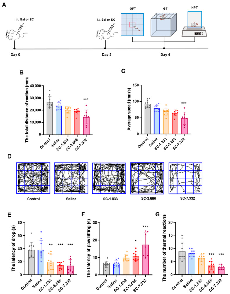
Source: Wang J et al. · Pharmaceutics · CC BY
Original figure and English caption ↗
Repeated exposure accentuates dose differences
Observations include tail twisting, hindlimb rigidity or weakness and abnormal movement. Some low-exposure responses recover quickly; higher groups recover more slowly and include deaths. No animals survive the first administration in the highest-exposure group.
For example, at 7.332 micrograms per gram body weight, six of the initial twenty mice die after the first exposure, and twelve cumulatively after the second. At 3.666 micrograms per gram, two deaths occur after repeated exposure. Repetition can markedly worsen outcomes.
Deaths emerge at an intermediate exposure after repeated dosing, while cumulative mortality increases in another higher group. Repetition matters alongside amount: temporary recovery after one exposure does not ensure the same response to the next.
Early marker reductions precede increases in selected regions
At ten and sixty minutes, c-Fos activity markers differ across regions. Early counts decrease in prelimbic cortex, or PrL, and infralimbic cortex, or IL. Later counts rise in PrL and secondary motor cortex, or M2.
PrL population calcium recordings similarly show a smaller signal area during the first ten minutes after the second exposure and a larger area at sixty to seventy minutes. Marker and signal time courses agree in direction, revealing dependence on the observation window.
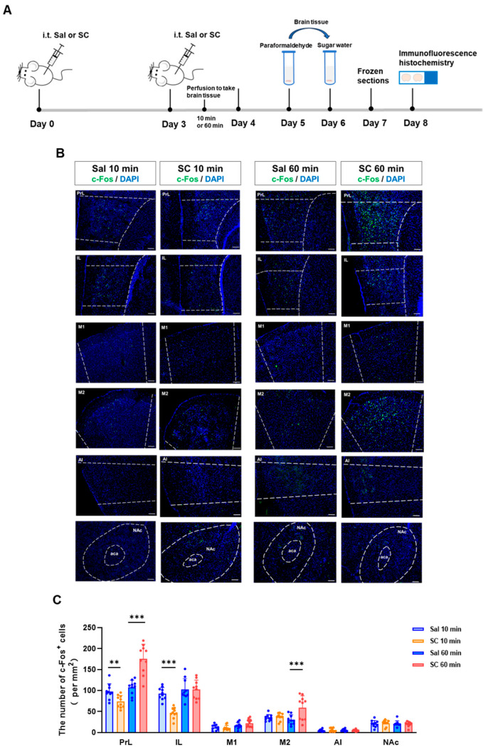
Source: Wang J et al. · Pharmaceutics · CC BY
Original figure and English caption ↗
One region shows two distinct response windows
Calcium signals report population activity while also reflecting cellular calcium processes. Early weakening and later enhancement in PrL motivate attention to both activity and ion environments across multiple time points.
Citrate’s capacity to bind metal ions motivates the authors’ calcium-homeostasis hypothesis. How do the biphasic fluorescence responses relate to citrate distribution, ion binding and neural activity? The signals define the temporal pattern that a mechanism must explain.
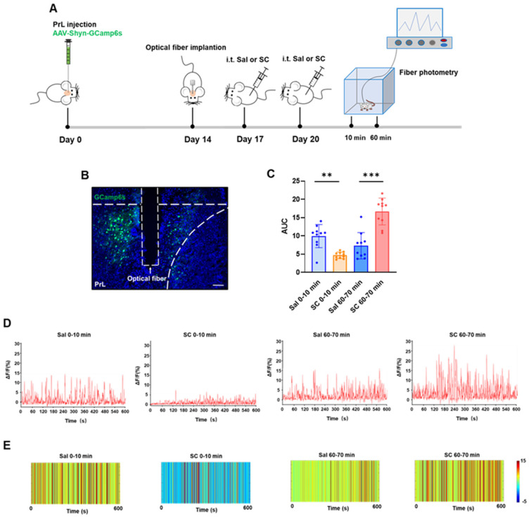
Source: Wang J et al. · Pharmaceutics · CC BY
Original figure and English caption ↗
Transient rabbit abnormalities add functional and tissue observations
In rabbits of both sexes, no evident abnormalities are observed in the two lower groups. Higher exposures produce arched posture, rigidity, tremor and struggling, with signs resolving within one to two hours. Reported CSF calcium and magnesium changes at higher exposure add an ion-environment clue.
No evident structural injury appears in examined injection-site tissue despite functional abnormalities. This animal study brings movement, sensory responses, transient neural signals and tissue morphology into formulation evaluation; functional responses and local tissue morphology describe different aspects of the response under these experimental conditions.
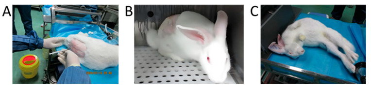
Source: Wang J et al. · Pharmaceutics · CC BY
Original figure and English caption ↗