Bringing a few cells out of a crowded background: how Tri-M combines sparsity with brightness
Three density levels, clear cellular processes and observations across months: a mouse study tests how sparse labeling can remain bright and withstand time.
Cell bodies, dendrites and axons overlap throughout brain tissue. Making every cell fluorescent does not always improve visibility: a fine process can quickly disappear among neighboring signals. Sparse labeling takes a different approach, illuminating only a few cells while giving them clear outlines. But can researchers choose the density, and will the image grow crowded over months? A 2026 Advanced Science study of Tri-M addresses both questions, demonstrating three labeling levels and following how different versions change with time.
Tri-M1 creates three density levels that remain stable over three to six months in the tested cortical population. Tri-M2 reaches closer to a single-cell scale, while the number of visible cells rises with time.
Contents · 7 sections
Fewer labeled cells can reveal more
Sparse labeling aims to make a few cells bright rather than dim the entire field. Cell bodies are only the beginning: long axons and small dendritic spines also need sufficient signal. Simply lowering expression can lose detail, while waiting longer can increase both brightness and the number of visible cells.
Tri-M separates the demands by first determining which cells enter an expression state, then allowing selected cells to retain clear outlines. Cell identity, location and labeling probability shape the image, giving density and brightness room to work together.
A probability switch sets the density
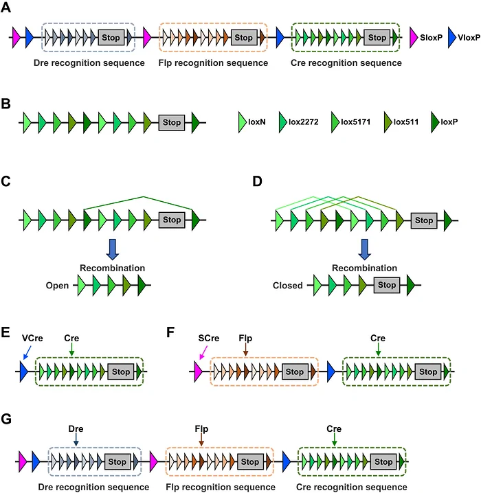
The design uses competition among intracellular recombination outcomes: most cells remain off, while a minority turn on. Different combinations create one, two or three probability gates and three density levels. Once established, the state is less likely to return to the original competition, providing the basis for sustained sparsity.
Tunability means choosing among density modes in advance. By combining the module with existing cell-type resources, the study examined expression in several brain regions. A shared probability framework could therefore reveal different populations at different densities.
Source: Tang X et al. · Advanced science (Weinheim, Baden-Wurttemberg, Germany) · CC BY
Original figure and English caption ↗
Three levels remain distinct months later
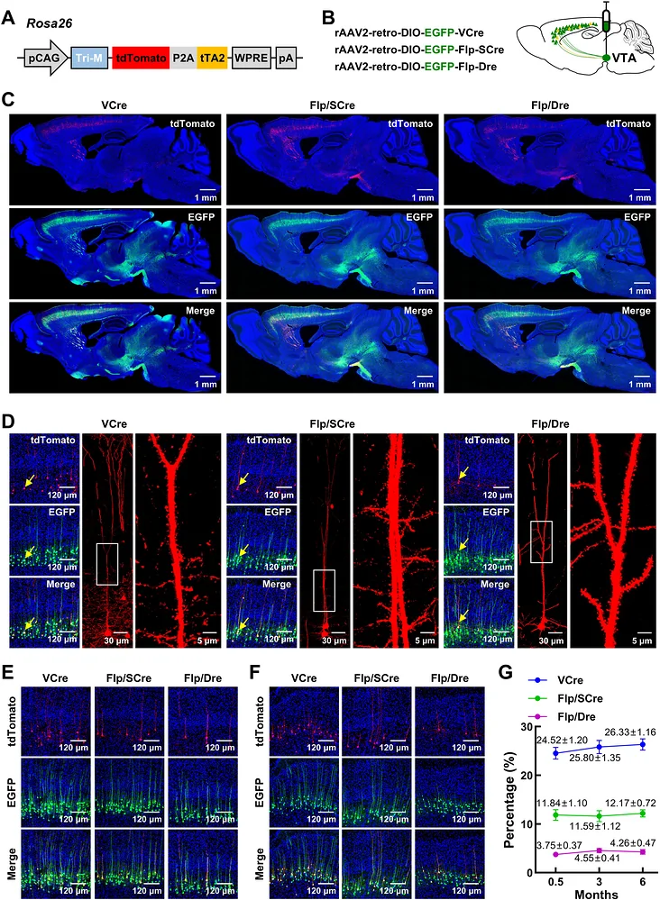
The initial demonstration examined layer-V excitatory cortical neurons projecting to ventral tegmental area. Green labeling identified the relevant experimental population, while red tdTomato showed its Tri-M1-positive subset. Red cells represented approximately24.5%,11.8% and3.8% of green cells across the three modes, producing clearly separated levels.
Selected cells retained visible axons and dendritic spines as well as cell bodies. The three density levels remained stable at three and six months, while controls without the relevant recombination condition showed no red cells. Time became a test of lasting image clarity rather than merely a wait for brighter signals.
Source: Tang X et al. · Advanced science (Weinheim, Baden-Wurttemberg, Germany) · CC BY
Original figure and English caption ↗
Across different brain-cell populations
The team also observed graded labeling in dentate granule cells, visual-cortical PV-positive cells and basal-forebrain cholinergic neurons. Despite their different shapes and networks, a shared framework brought a small number of cells out of the background.
Astrocytes add another question: genetic states can pass to daughter cells during division, making the timing of labeling important. A temporally controlled CA1 demonstration produced three density levels. Sparse images thus depend on probability as well as developmental and proliferative state.
Beyond visibility, a functional response
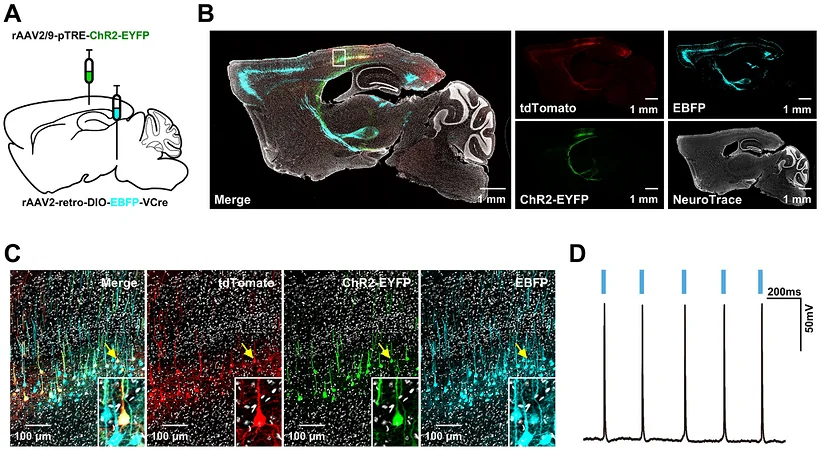
The researchers next asked whether visible cells could carry a functional component. Tri-M1-labeled sensory-cortical cells expressing a light-sensitive protein showed corresponding light-evoked action potentials. Morphological visibility and functional responsiveness came together in the same small population.
The result opens a path from identifying a cell to observing its response and asking what it contributes to a network. The study establishes cellular responsiveness, with behavioral roles left for future questions.
Source: Tang X et al. · Advanced science (Weinheim, Baden-Wurttemberg, Germany) · CC BY
Original figure and English caption ↗
Near a single-cell scale, time becomes a variable
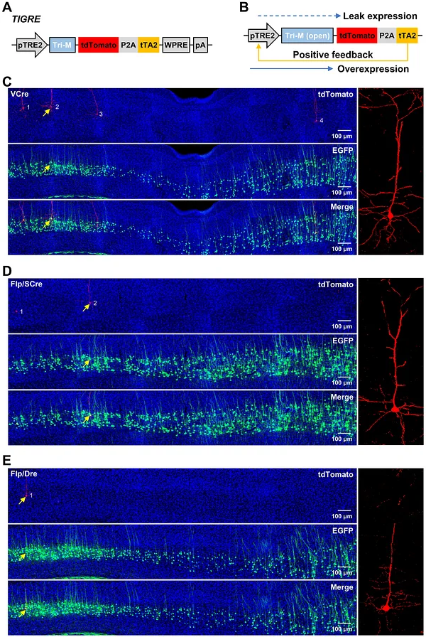
Tri-M2 combines limited initial expression with subsequent signal amplification to reduce the number of visible cells further. Its three levels were approximately7,3 and0.7 per thousand; sometimes only one red cell appeared in a section. Fewer cells with clear outlines make an individual axon or dendritic arbor easier to follow.
The sparser version follows a different trajectory: more Tri-M2 cells become visible with time, which the authors associate with accumulated opportunities for weak initial expression. At six months, the lowest level remained around1 per thousand—very sparse, but changed. The versions therefore offer different strengths: long-term stability and closer-to-single-cell resolution.
Source: Tang X et al. · Advanced science (Weinheim, Baden-Wurttemberg, Germany) · CC BY
Original figure and English caption ↗
Clear images reveal a set of trade-offs
The study brings several determinants of a clear image into one framework: cell identity, number, signal strength and observation time. Population studies and single-cell tracing may call for different densities, while short observations and months-long tracking may favor different versions.
The evidence concerns neurons and astrocytes in mouse brain. Other tissues, species and long-term applications remain to be explored, alongside the complexity of the required combinations. Tri-M makes sparse yet bright labeling a set of choices, opening more room to observe cellular detail.