A new color for acetylcholine reveals coordinated brain signals
Red GRAB sensors enable simultaneous imaging of acetylcholine and other neuromodulators, revealing distinct dynamics during learning, sleep and sound responses.
Brain signals can resemble an ensemble: one part begins as another follows or shifts to a different rhythm. Listening to only one part makes their coordination difficult to understand. Acetylcholine participates in many neural functions, yet its relationship with other neuromodulators calls for simultaneous observation. A study published in Nature Neuroscience on June 16, 2026 develops red fluorescent GRAB acetylcholine sensors. Paired with green sensors, the red channel permits two neurochemical signals to be compared on the same behavioral timeline in mice.
The high-affinity red sensor rACh1h reports acetylcholine dynamics across brain regions and pairs with green neuromodulator sensors. Learning, sleep transitions and cortical sound responses reveal related but distinct temporal and spatial profiles.
Contents · 7 sections
An added color opens a simultaneous view
Acetylcholine acts within a broader neuromodulatory environment. Recording one signal alone can miss its timing relative to another. A red sensor leaves the green channel available to report dopamine, serotonin or norepinephrine alongside acetylcholine.
Both signals can now be measured during the same sound, sleep episode or brief behavioral event. Shared context makes it possible to compare co-occurrence, timing and divergent changes. The question expands from when one transmitter changes to how several signals vary together.
A red signal must still identify acetylcholine
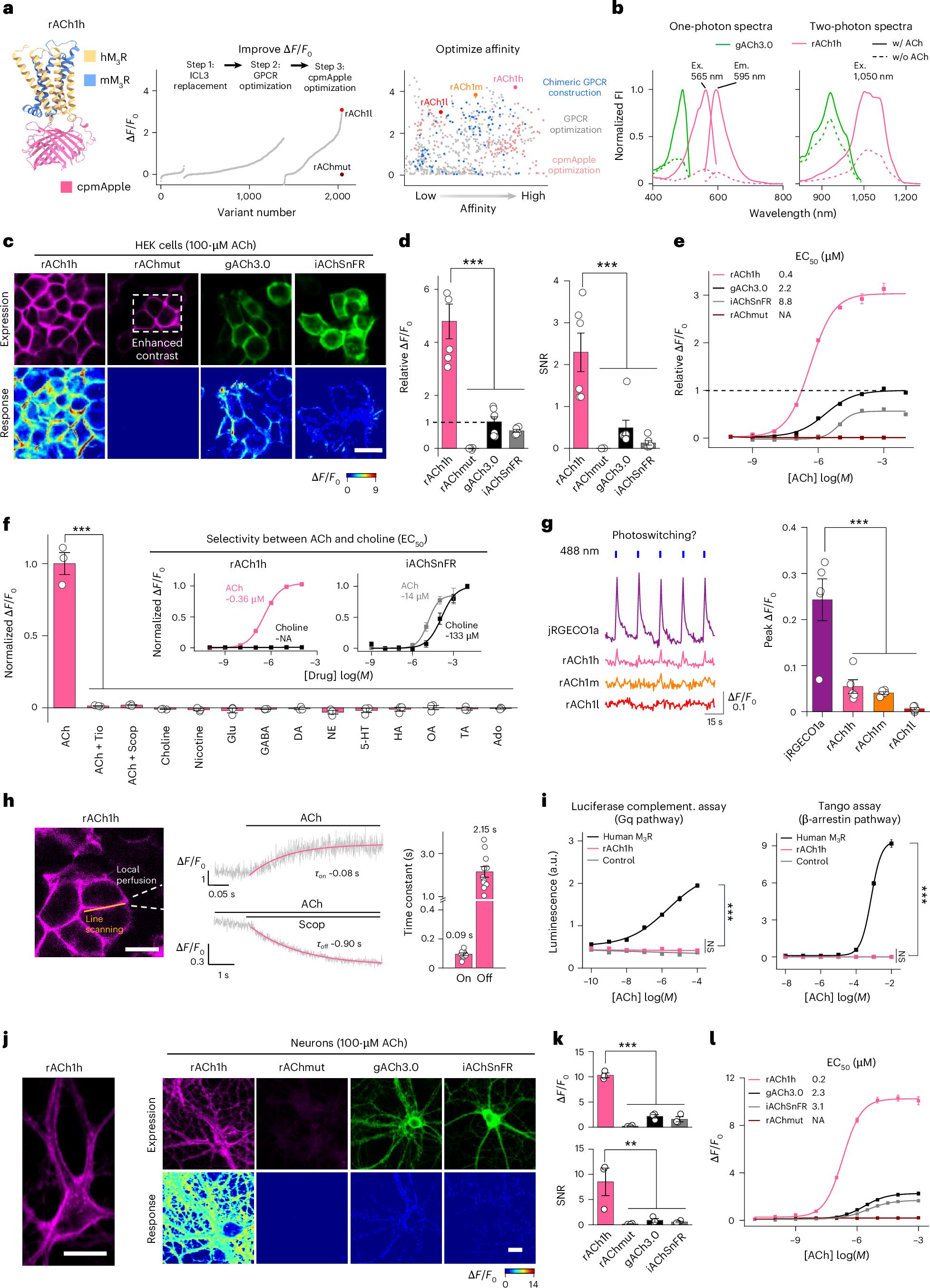
The sensor series includes variants with different affinities. The high-affinity rACh1h responds at lower acetylcholine concentrations and shows larger fluorescence changes and higher signal-to-noise ratios in the tested cell and cultured-neuron comparisons. Acetylcholine binding is translated into a measurable brightness change.
Controls support the interpretation: other tested compounds did not produce the same response, receptor antagonists suppressed acetylcholine-associated signals, and blue-light-induced changes were smaller than in the red calcium-indicator comparator. Downstream coupling assays also lacked the marked responses seen with the wild-type receptor, supporting its role as a reporter under the tested conditions.
Source: Shu Xie et al. · Nature Neuroscience ·
Original figure and English caption ↗
From cellular brightness to endogenous brain release
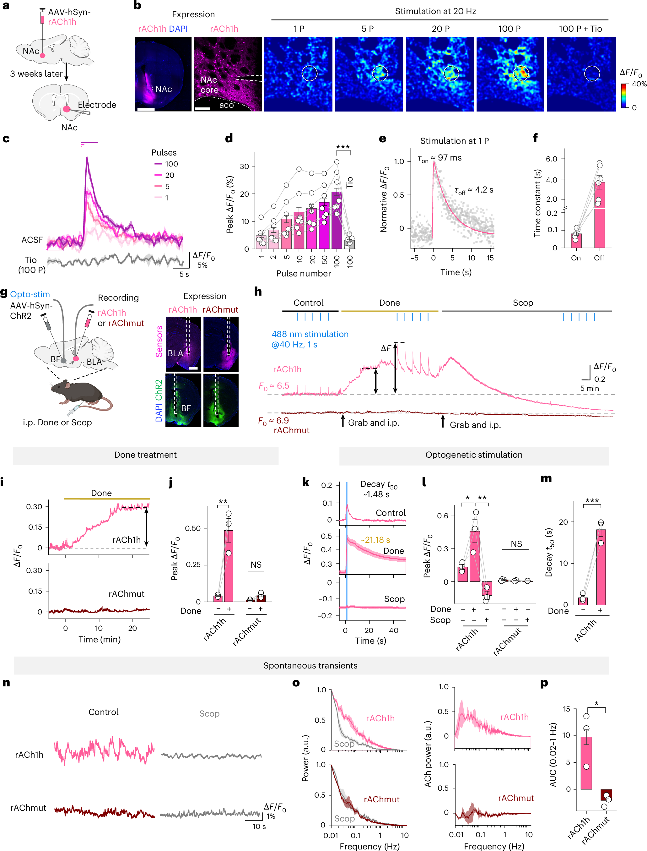
In accumbal brain slices, stimulation-evoked fluorescence increased with pulse number and was strongly reduced by a receptor antagonist. A single pulse also produced a rapid rise followed by decay, making endogenous release dynamics observable.
In the mouse amygdala, activating basal-forebrain cholinergic inputs evoked red signals. Inhibiting acetylcholine breakdown increased responses and slowed their decay, while receptor antagonism reduced them. A response-deficient mutant did not show the same pattern, providing complementary validation of the relation between fluorescence, release and clearance.
Source: Shu Xie et al. · Nature Neuroscience ·
Original figure and English caption ↗
Two neuromodulators respond to a learned cue
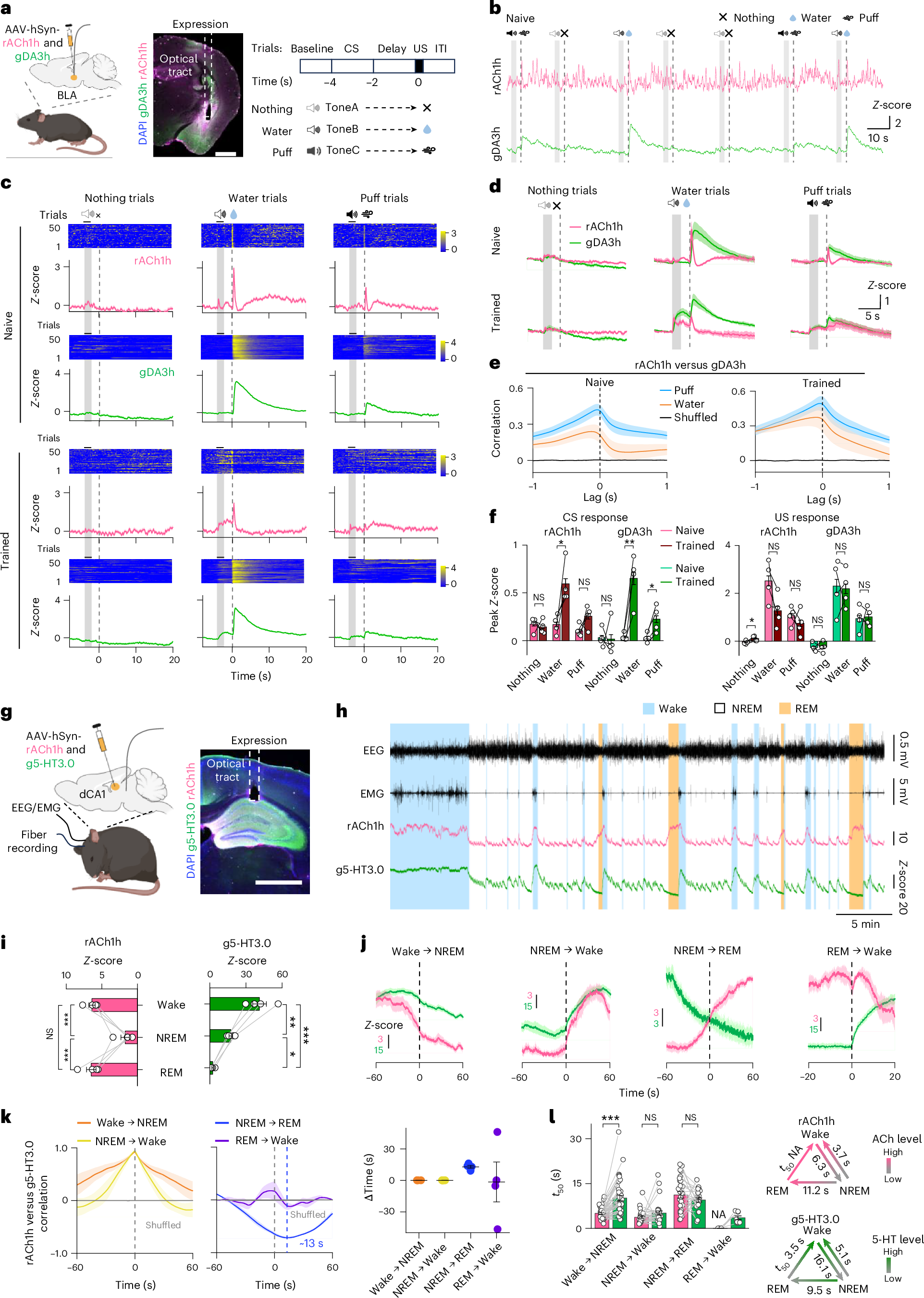
Simultaneous acetylcholine and dopamine recordings in the basolateral amygdala followed learning about sounds predicting water, air puff or no outcome. After training, responses to the water-predicting cue strengthened in both channels, while dopamine responses also strengthened for the puff-predicting cue.
Changes were not uniform across conditions or time windows. The acetylcholine comparison for the puff cue and several outcome-period comparisons were not significant. The two-color view preserves these distinctions: learning affects neuromodulatory cue responses, but the channels do not simply report identical dynamics.
Two signals diverge at entry into REM sleep
In dorsal CA1, acetylcholine and serotonin reporters revealed different profiles across wakefulness, NREM and REM sleep. Acetylcholine signals were lower in NREM and higher in wakefulness and REM. Serotonin signals decreased from wakefulness to NREM and further in REM.
During NREM-to-REM transitions, acetylcholine rose as serotonin fell. During wake-to-NREM transitions, both decreased with different temporal profiles. Simultaneous recordings describe coordination and timing; establishing whether one change drives another requires causal investigation.
Source: Shu Xie et al. · Nature Neuroscience ·
Original figure and English caption ↗
A sound leaves distinct cortical chemical maps
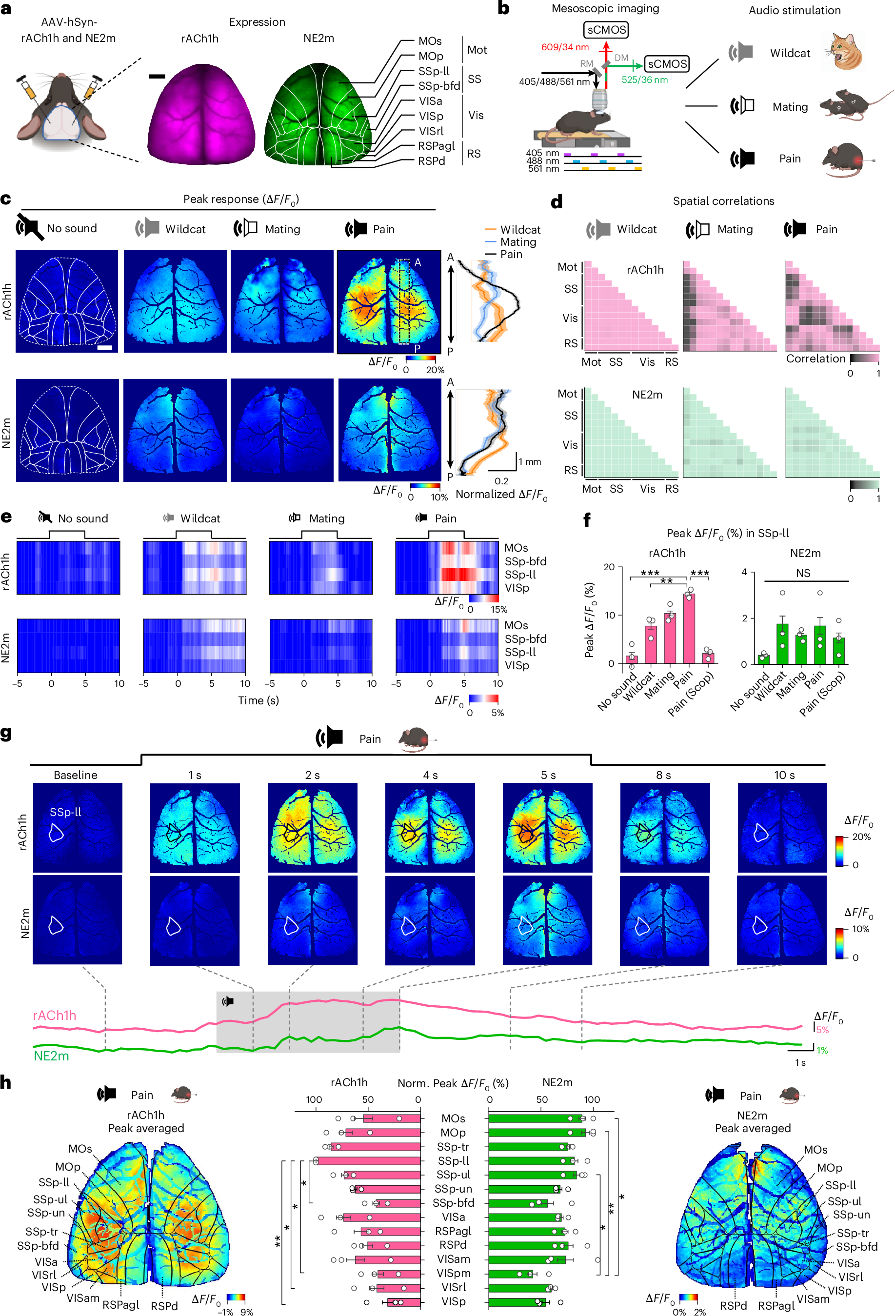
Across the dorsal cortex, acetylcholine and norepinephrine responses also differed spatially. Imaging during animal-sound playback showed regionally stronger acetylcholine responses to pain vocalizations, with a spatial pattern distinct from norepinephrine.
In lower-limb primary somatosensory cortex, acetylcholine responses were stronger for pain vocalizations in the reported comparisons, whereas the corresponding norepinephrine comparison across conditions was not significant. These are sound-evoked transmitter measurements; they do not establish the animal’s subjective experience or assign one function to every responsive region.
Source: Shu Xie et al. · Nature Neuroscience ·
Original figure and English caption ↗
A shared timeline changes the questions we can ask
The advance concerns comparison as well as color. Learned cues in the amygdala, sleep transitions in the hippocampus and sound responses across cortex can be viewed alongside another neurochemical signal in a shared context.
Affinity, response and decay kinetics, recording location and behavioral conditions shape the observed traces. Simultaneous imaging does not itself establish causal interactions, but it enables more precise questions about coordination, divergence and temporal ordering. Tool development thus brings previously separate measurements into a common view for mechanistic investigation.