Why does strengthening thalamic–cingulate responses make mice more sensitive?
Patterned stimulation strengthens pathway responses in freely moving male mice; low-frequency stimulation reduces potentiation and induced hypersensitivity without matching anxiety-like changes.

The touch may be the same, yet the brain can respond more strongly. If an input carrying sensory information is potentiated, how long does sensitivity last—and can it be reduced? Thalamus links sensory input with cortical processing, while anterior cingulate cortex, or ACC, contributes to pain-related processing. The strength of the connection offers a route into understanding persistent hypersensitivity. A Molecular Pain study examines medial thalamic, or mTH, input to ACC in freely moving adult male mice. One patterned stimulation strengthens pathway responses and increases mechanical and heat sensitivity; a different pattern reduces both potentiation and induced hypersensitivity.
TBS produces persistent mTH–ACC potentiation, retained at six weeks in some animals. Sensory hypersensitivity occurs over a week without significant anxiety-like changes; LFS reduces TBS potentiation and hypersensitivity.
Contents · 6 sections
Selected thalamic input can be potentiated
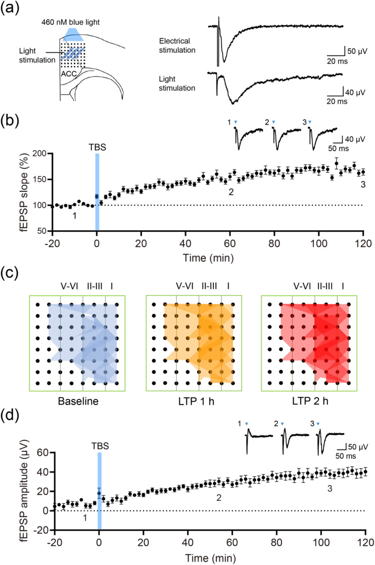
ACC has multiple inputs. Selecting light-responsive thalamic axons and recording field excitatory postsynaptic potentials isolates this route more closely. TBS produces potentiation lasting at least two to three hours, including when responses are tested optically.
Previously unresponsive recording channels acquire persistent signals. Potentiation increases existing responses and extends detectable activity to previously silent recording positions, changing the response distribution.
Field potentials capture local synaptic responses, whose slopes track input efficacy. Comparing the same input before and after induction reveals stronger responses to comparable tests. Persistent potentiation retains effects of activity history, motivating its study in lasting hypersensitivity.
Source: Lin S et al. · Molecular pain · CC BY
Original figure and English caption ↗
From hours to weeks
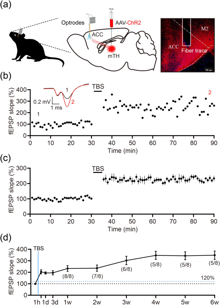
In freely moving mice, TBS rapidly raises responses after a stable baseline and maintains them over the first hour. Without induction, test responses are relatively stable across days, supporting the link with stimulation history.
All eight longitudinal animals show potentiation initially. Five retain it at six weeks while three return to normal. A brief induction can leave weeks-long changes, with differing recovery across animals.
Source: Lin S et al. · Molecular pain · CC BY
Original figure and English caption ↗
Potentiation accompanies greater stimulus sensitivity
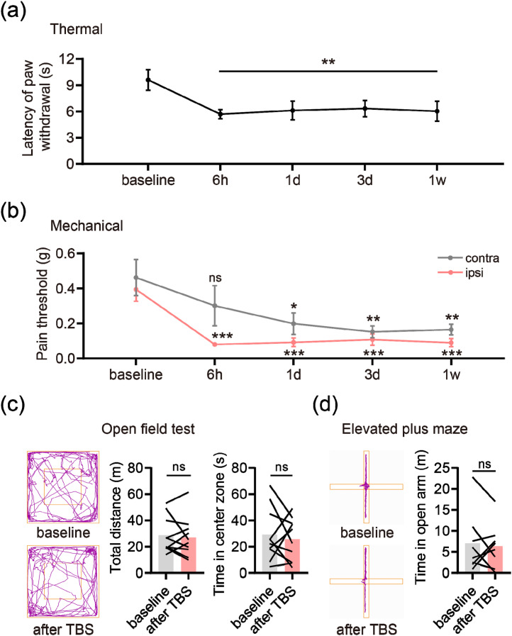
TBS shortens heat-response latency through one week. Mechanical thresholds fall more prominently ipsilaterally early on; the six-hour contralateral comparison is nonsignificant, with later decreases from day one to one week. Changes have temporal and lateral distinctions.
Open-field distance and center duration and plus-maze measures do not significantly change. Greater mechanical/heat sensitivity coexists with comparable tested locomotor and anxiety-like outcomes.
These TBS experiments do not first induce peripheral injury. Altering central input activity is sufficient for later sensory hypersensitivity, separating the contribution of central potentiation for comparison with inflammatory changes.
Source: Lin S et al. · Molecular pain · CC BY
Original figure and English caption ↗
Peripheral inflammation also enhances the input
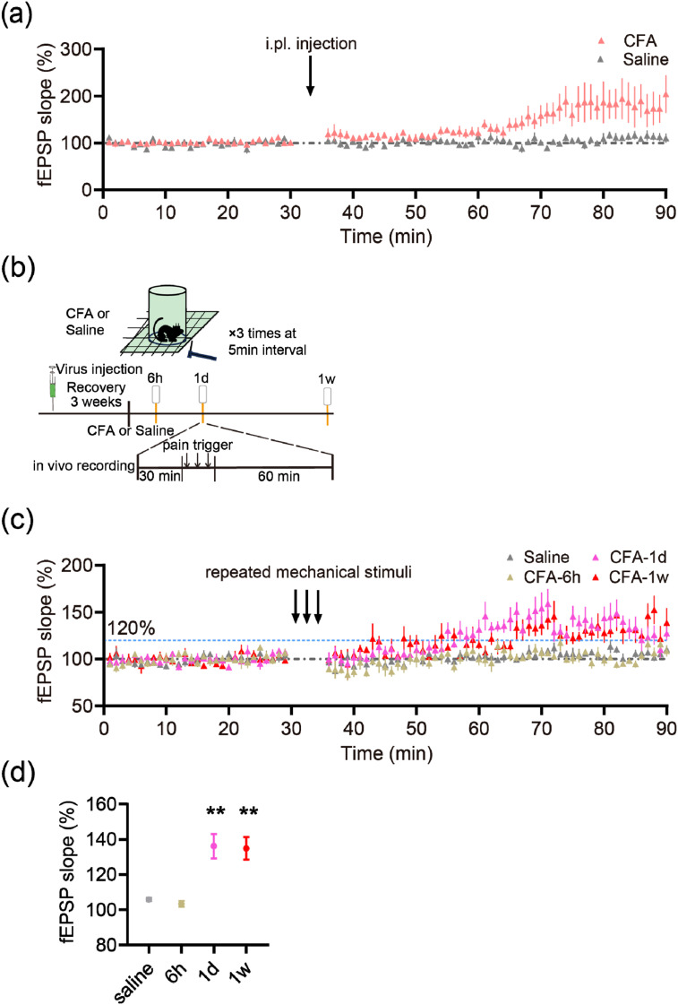
Following plantar CFA inflammation, optically tested mTH–ACC responses rise early. Repeated mechanical stimuli later enhance model responses more than saline controls. Peripheral tissue state relates to central input responsiveness beyond artificial induction.
The original figure shows clearer later day-one and week-one changes than at six hours. Inputs reach a pathway whose state changes with time, producing different subsequent response strengths across stages.
Source: Lin S et al. · Molecular pain · CC BY
Original figure and English caption ↗
Low frequency reduces potentiation
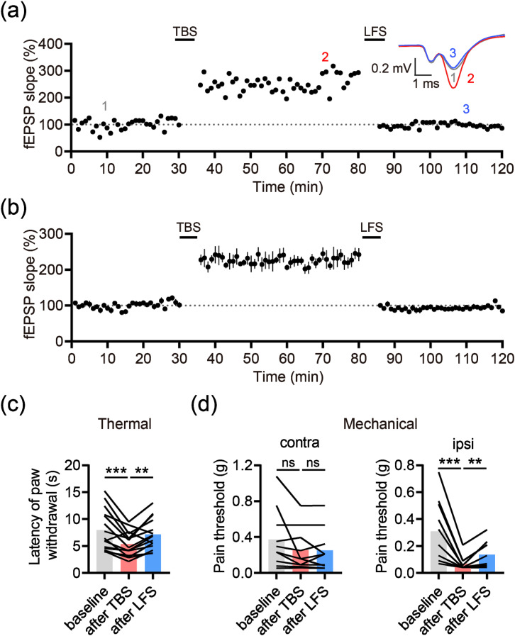
LFS one hour after TBS returns responses toward baseline and significantly reduces the potentiated state. The route supports opposite plastic changes under different activity patterns.
Heat and ipsilateral mechanical hypersensitivity improve, while contralateral changes are nonsignificant. This rescue concerns TBS-induced hypersensitivity, linking reduced physiological potentiation with sensory outcomes.
Source: Lin S et al. · Molecular pain · CC BY
Original figure and English caption ↗
Sensory modulation has adjustable connection strength
Adult male-mouse results connect potentiation and hypersensitivity within one input route. Weeks-long physiology, week-long behavior and opposite modulation reveal persistent yet adjustable strength. Sensory reception depends partly on the pathway’s activity history.