Why does sleep loss prolong post-injury sensitivity? A brainstem–thalamus–prefrontal route
A male-mouse incision study tracks recovery over time: upstream connections affect both wakefulness and sensitivity, while a prefrontal output separates the two.
Wounds and nervous-system sensitivity each have a recovery timetable. What happens to that timetable when sleep is lost? A Behavioral and Brain Functions study compares mechanical, cold and thermal responses after incision in male mice, following connections from locus coeruleus through paraventricular thalamus to prefrontal cortex. Sleep loss prolongs sensitivity, but the network reveals a division of labor: some connections change both wakefulness and stimulus responses, whereas one output changes sensitivity without a clear change in wakefulness.
PVT links a brainstem arousal-related input with a cortical output involved in persistent post-injury sensitivity; sleep state and pain-like responses can vary together or separately.
Contents · 6 sections
Why does sensitivity linger after the same injury?
Mice with incision alone approach baseline later in the study, while mice with incision and sleep loss remain more responsive. Stronger mechanical and cold responses and shorter thermal-response latency trace delayed recovery. Following observable responses over time reveals changes that a single-day measurement could miss.
A shorter period of sleep loss does not significantly alter baseline thresholds in uninjured mice; longer exposure produces baseline sensitization. Sleep changes therefore have different consequences in an injured state. The question concerns both stronger responses and why sensitivity remains during the expected recovery period.
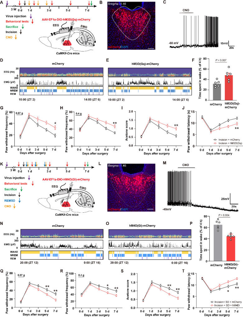
PVT activity affects wakefulness and recovery
Incision combined with sleep loss increases activity-related labeling in PVT glutamatergic cells. Raising their activity increases wakefulness and sustains stronger mechanical, cold and thermal responses later after incision. Lowering activity reduces wakefulness and persistent sensitivity in the sleep-loss model. PVT activity relates bidirectionally to both outcomes.
The paired changes make PVT a meeting point and raise a question: does altered sensitivity follow sleep state, or travel through another output? A thalamic node can send information to different targets, so changing its overall activity may combine several contributions. Comparing inputs and outputs exposes its network roles.
Source: Li L et al. · Behavioral and brain functions : BBF · CC BY-NC-ND
Original figure and English caption ↗
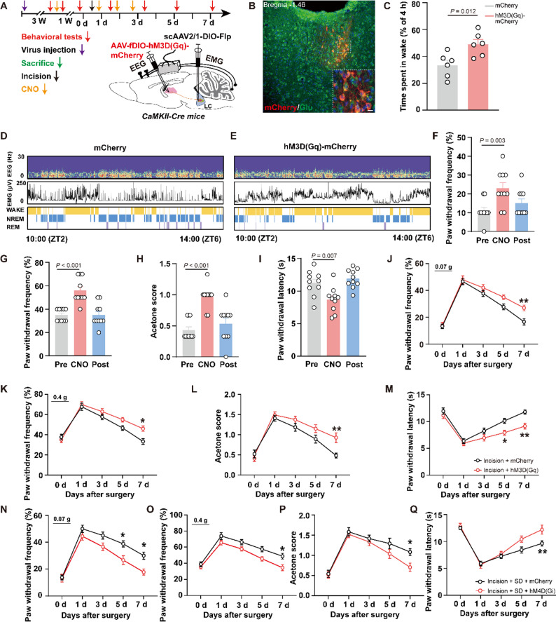
Locus coeruleus links arousal with persistent sensitivity
Locus coeruleus (LC) participates in arousal regulation. Its TH-positive cells show more activity-related labeling in the incision-plus-sleep-loss model, and tracing supports LC input to PVT. Increasing this input strengthens PVT calcium signals and stimulus responses; reducing it weakens the signals and improves sensitivity measures.
Increasing activity in the associated population raises wakefulness, reduces sleep and delays recovery of post-incision responses; reducing it shifts outcomes oppositely. Brainstem arousal-related signals connect with thalamic recovery trajectories. TH identifies a catecholamine-associated population; the complete transmitter–receptor relay remains unresolved, while the activity–state–behavior relationship is evident.
Source: Li L et al. · Behavioral and brain functions : BBF · CC BY-NC-ND
Original figure and English caption ↗
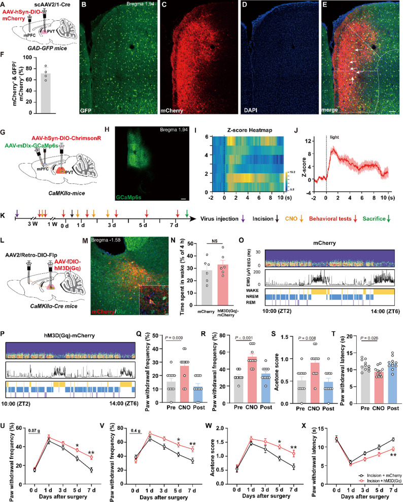
Greater sensitivity without a clear rise in wakefulness
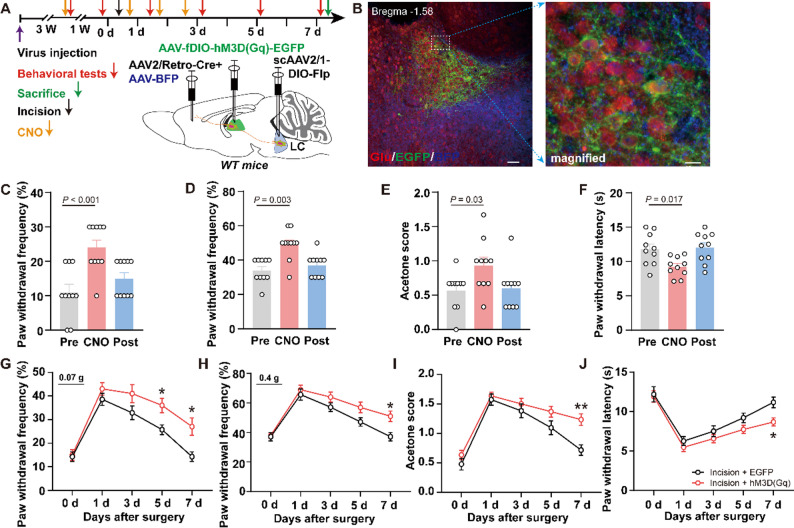
PVT output reaches medial prefrontal cortex (mPFC), with substantial labeling associated with GABAergic cells. Increasing PVT-derived input strengthens calcium signals in these cortical cells, enhances mechanical and cold responses, shortens thermal latency and prolongs post-incision sensitivity. The route now extends from brainstem state regulation toward cortical stimulus processing.
Wakefulness during the measured period does not change significantly. Sensitivity shifts without an equivalent movement in sleep state. Reducing this output leaves baseline thresholds largely unchanged in uninjured mice but improves outcomes with incision and sleep loss. The output’s contribution depends on both its target and the animal’s state.
Source: Li L et al. · Behavioral and brain functions : BBF · CC BY-NC-ND
Original figure and English caption ↗
Input and output meet in a thalamic population
The study examines PVT populations associated with LC input and mPFC output. Increasing their activity enhances baseline responses and delays post-incision recovery; reducing it attenuates persistent responses in the sleep-loss model. The results join the nodes in a framework of multiple connections, placing thalamus at the intersection of input and output effects.
The framework helps explain the division of labor along the route. Upstream changes closely accompany wakefulness, whereas the cortical output separates sensitivity from arousal. The populations may have other connections, but these findings give recovery a spatial organization: persistence can be related to particular nodes and targets.
Source: Li L et al. · Behavioral and brain functions : BBF · CC BY-NC-ND
Original figure and English caption ↗
Sleep and sensitivity share a network with distinct trajectories
In this male-mouse incision study, recovery extends from events around the wound to connections in the brain. The clearest finding is that sleep and sensitivity can change together yet separate at one output. Recovery timing becomes part of understanding network function: the animal’s state and the signal’s destination influence when sensitivity subsides.