Why does facial touch provoke stronger responses? A hypothalamic CRF link to the trigeminal brainstem
A mouse study connects hypothalamic CRF populations, brainstem sensory input and CRFR2, outlining a descending route involved in facial hypersensitivity.
Why does the nervous system sometimes respond more strongly to the same light touch? Facial signals enter the trigeminal system, while brain state may alter how they are processed. An Advanced Science study examines hypothalamic paraventricular nucleus together with spinal trigeminal nucleus caudalis in the brainstem. In a nitroglycerin migraine-like mouse model, hypothalamic CRF populations become more active and local brainstem signals and sensory responses change. A descending route from a state-regulating region enters the story of hypersensitivity.
PVN CRF-population activity relates bidirectionally to model hypersensitivity; local SP5C CRF enhances cellular responses, with clearer CRFR2-related effects than CRFR1 effects under the tested conditions.
Contents · 6 sections
Descending signals can regulate brainstem input
Spinal trigeminal nucleus caudalis (SP5C) processes head and facial nociceptive input in the brainstem. Hypothalamic paraventricular nucleus (PVN) contains diverse neuropeptide populations involved in bodily states. Linking the regions raises a specific question: can hypothalamic signals increase brainstem responsiveness to the same touch?
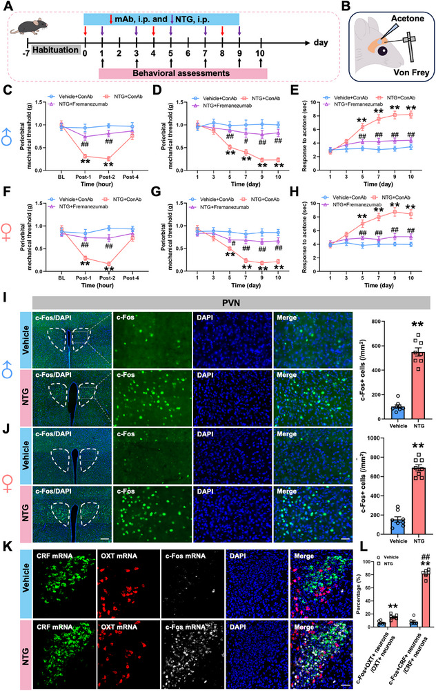
The model’s time course provides context. Mechanical sensitivity after one nitroglycerin exposure is transient, whereas repeated exposure sustains lower periorbital thresholds and longer cold responses. Main figures show baseline phenotypes in both sexes, and an anti-CGRP comparison attenuates responses. Transient and persistent sensitivity become distinct states to compare.
Activity-related labeling rises in PVN, with stronger recruitment of CRF than the tested OXT population. Cells in one region do not change uniformly. The population difference focuses attention on CRF: how does its activity relate to sensitivity, and where can it send signals?
Source: Bian J et al. · Advanced science (Weinheim, Baden-Wurttemberg, Germany) · CC BY
Original figure and English caption ↗
Sensitivity changes with CRF activity
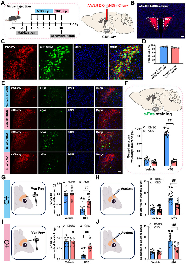
PVN CRF calcium activity and responses to light touch increase in the model. Reducing activity raises periorbital mechanical thresholds and weakens cold responses; increasing it produces mechanical and cold sensitivity without the same established model state. Opposite activity changes yield corresponding behavioral shifts, supporting functional involvement.
Male and female population results in the main figures point in the same direction, while baseline controls do not change like the model. The effect depends on state. Mechanical and cold readouts connect a neuropeptide population with specific behaviors and motivate the search for its downstream target.
Source: Bian J et al. · Advanced science (Weinheim, Baden-Wurttemberg, Germany) · CC BY
Original figure and English caption ↗
From hypothalamus to a trigeminal sensory node
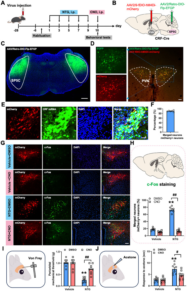
Tracing supports PVN CRF connections with SP5C, where most labeled downstream cells carry glutamatergic markers. Reducing SP5C-projecting PVN CRF activity improves mechanical measures. Some cold comparisons also change, although overall intervention and interaction tests are nonsignificant; the mechanical evidence is clearer.
Associated interventions reduce activity labeling in anterior cingulate and insular cortex. PVN populations projecting to those cortical areas overlap little with SP5C-projecting cells, suggesting possible indirect effects on higher regions. Descending signals and ascending sensory processing meet across several regions rather than operating in an isolated pair.
Source: Bian J et al. · Advanced science (Weinheim, Baden-Wurttemberg, Germany) · CC BY
Original figure and English caption ↗
CRF increases responsiveness to input
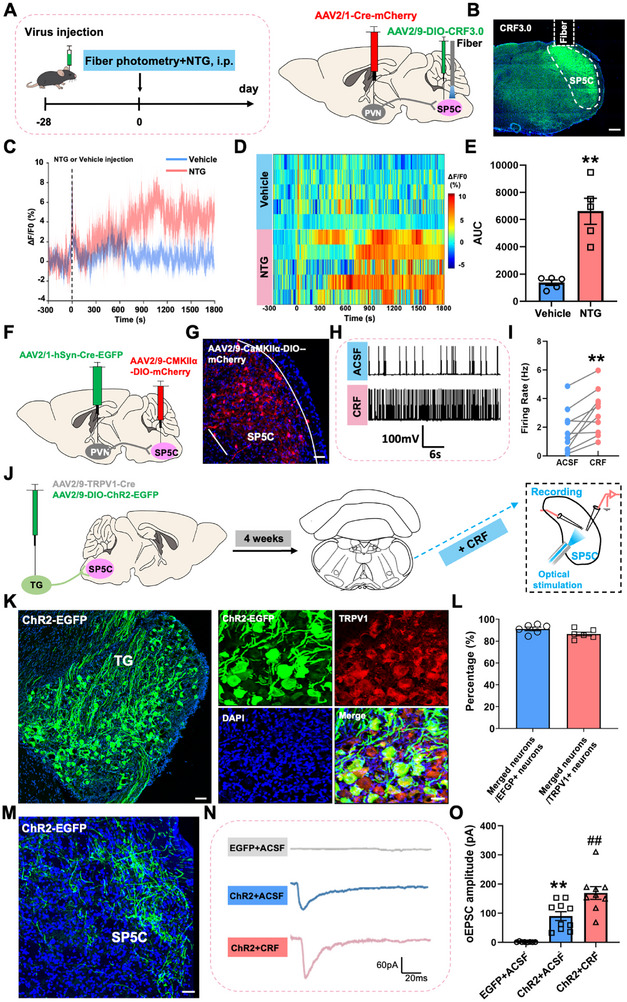
Local CRF-associated sensor signals rise in SP5C under nitroglycerin. In ex vivo observations, CRF increases firing in examined cells and the amplitude of evoked excitatory currents from trigeminal input. Cellular and input measures converge: the local network can respond more strongly when a signal arrives.
Source populations, local peptide signals and downstream responses provide evidence at different levels. A sensor measures local signaling without assigning every peptide source. Together, the observations support a model involving PVN-associated output and SP5C CRF regulation, centered on changed responsiveness to sensory input.
Source: Bian J et al. · Advanced science (Weinheim, Baden-Wurttemberg, Germany) · CC BY
Original figure and English caption ↗
The same peptide has different receptor-dependent effects
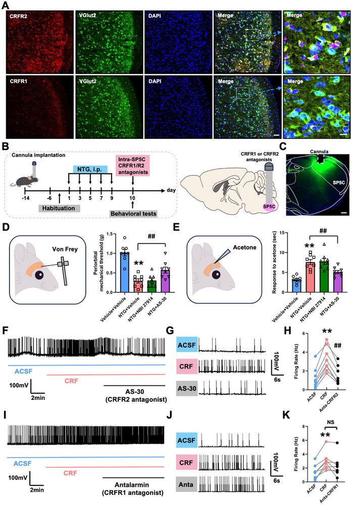
CRFR2 labeling appears in SP5C excitatory cells. CRFR2 blockade reduces model mechanical and cold responses and suppresses CRF-related firing enhancement. CRFR1 blockade shows no similarly clear benefit, and the overall related firing comparison is nonsignificant. Receptor identity becomes an important division of labor after peptide signals arrive.
The comparison narrows peptide effects to receptors at a local node. Rather than assigning one direction to all PVN outputs or CRF functions, the study finds clearer CRFR2-related effects here. Sensitization involves choices of source, target and receptor, each potentially shaping the final response to the same input.
Source: Bian J et al. · Advanced science (Weinheim, Baden-Wurttemberg, Germany) · CC BY
Original figure and English caption ↗
From cells to imaging, attention converges on the same regions
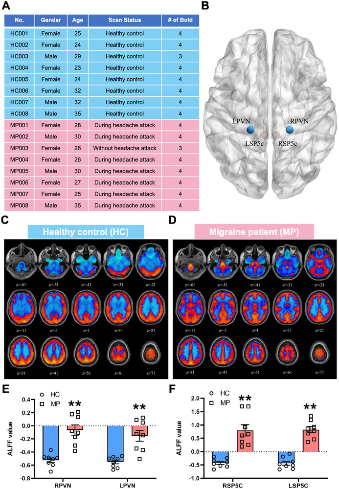
The study also compares imaging from eight participants with migraine and eight controls, finding regional low-frequency amplitude differences in PVN and SP5C; most patients were scanned during attacks. Imaging concerns regional measures, while mouse work follows cells and receptors. The observations draw attention to the same regions at different levels, with mechanistic evidence coming from the animal experiments.
The central insight is that facial sensitivity also has descending organization. As sensory input enters the brainstem, hypothalamic populations and local CRF receptors participate in regulating responses. Mechanical and cold changes in the migraine-like model gain a traceable route: how state signals reach a sensory node and change its responsiveness.
Source: Bian J et al. · Advanced science (Weinheim, Baden-Wurttemberg, Germany) · CC BY
Original figure and English caption ↗