How does brainstem processing amplify orofacial hypersensitivity after bite interference?
Trigeminal-input-associated parabrachial cells influence medullary and trigeminal stimulus responses, mechanical sensitivity and place avoidance in male rats.
Changing tooth contact can produce lasting masseter-region sensitivity in rats. After sensory information enters brainstem, processing is not simply one-way: ascending networks influence descending modulation and earlier sensory stages. A male-rat study in The Journal of Headache and Pain examines interactions among spinal trigeminal nucleus, or Sp5, lateral parabrachial nucleus, or LPBN, and rostral ventromedial medulla, or RVM. Manipulating Sp5-recipient glutamatergic-associated LPBN cells changes stimulus responses in both brainstem regions.
Activating Sp5-input-associated LPBN cells increases RVM ON-cell and Sp5 responses and induces mechanical hypersensitivity and place aversion. In the model, inhibition reduces neural responses, improves thresholds and produces preference for the inhibition-paired setting.
Contents · 7 sections
Input recipients and output cells differ
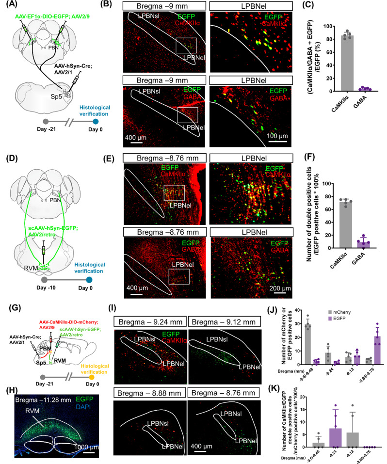
Most Sp5 cells projecting to LPBN carry the glutamatergic-associated CaMKIIα marker, with stronger ipsilateral and caudal terminal labeling. Approximately 86% of labeled Sp5-recipient LPBN cells carry CaMKIIα and 4% GABA; approximately 71% of RVM-projecting LPBN cells carry CaMKIIα.
The two links do not form a simple single-cell relay. Sp5-recipient LPBN cells are more caudal, RVM-projecting cells more rostral, with very little overlap. Information may require an intervening transfer within LPBN. Functional manipulation then asks whether the recipient population can still influence RVM.
Source: Mo S et al. · The journal of headache and pain · CC BY-NC-ND
Original figure and English caption ↗
Recipient activation strengthens ON-cell responses
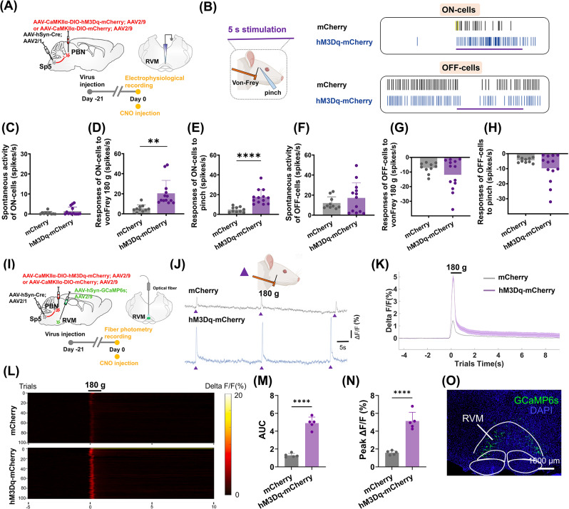
Activating Sp5-recipient glutamatergic-associated LPBN cells in nonmodel rats increases RVM ON-cell responses to orofacial mechanical and pinch stimuli during anesthetized recordings. Spontaneous ON firing and spontaneous or evoked OFF-cell activity do not significantly change.
RVM population calcium in another cohort shows larger stimulus-evoked peaks and areas. Single-cell and population findings align, although calcium samples a broader RVM population. Strengthening the upstream recipient group increases subsequent medullary stimulus responses.
Source: Mo S et al. · The journal of headache and pain · CC BY-NC-ND
Original figure and English caption ↗
Stimulus responses also increase in Sp5
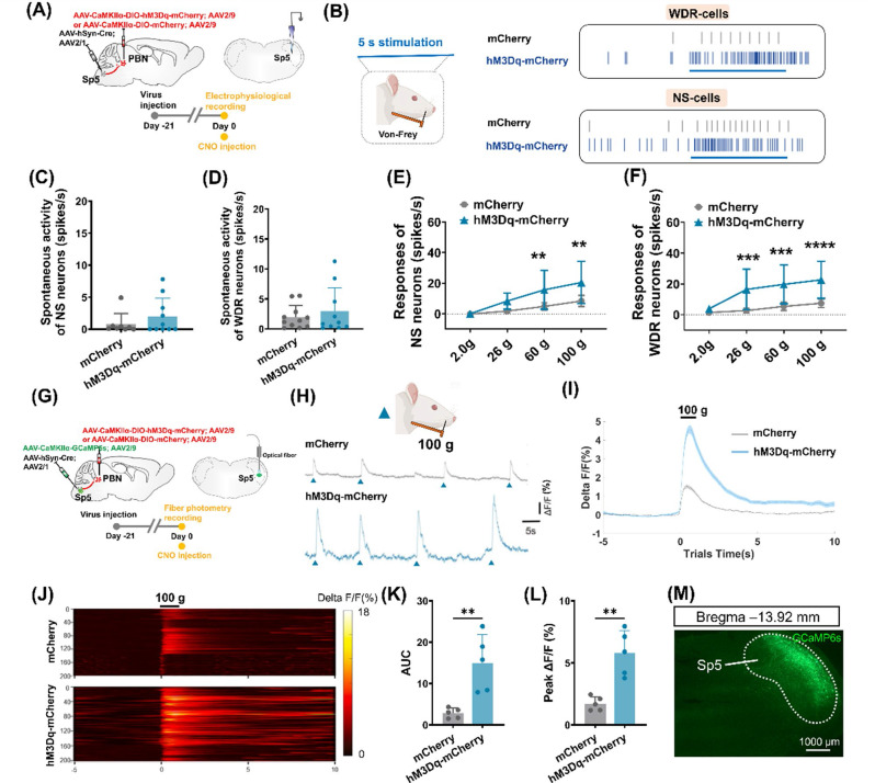
Sp5 is an early central processing site for orofacial information. LPBN activation enhances wide-dynamic-range, or WDR, responses to moderate and stronger stimuli, while nociceptive-specific, or NS, responses increase mainly at stronger intensities. Neither population shows significant spontaneous-firing changes.
Calcium in glutamatergic-associated Sp5 cells also increases. Manipulating LPBN recipients alters processing back at Sp5, consistent with interaction between ascending information and descending modulation. Hypersensitivity therefore involves more than peripheral input alone.
Source: Mo S et al. · The journal of headache and pain · CC BY-NC-ND
Original figure and English caption ↗
Lower withdrawal thresholds and place avoidance
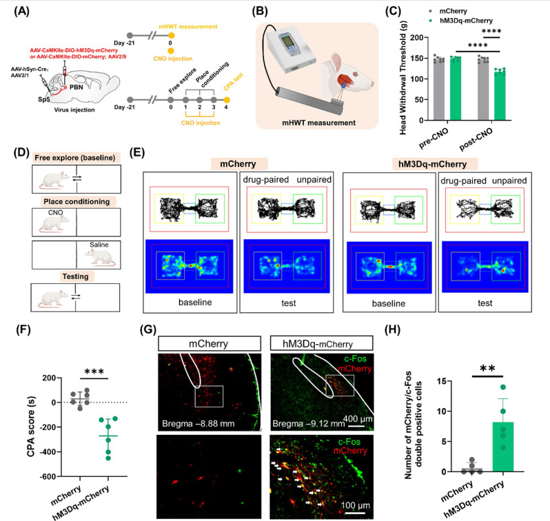
After recipient activation, the mechanical head-withdrawal threshold in the masseter region falls: less pressure evokes a response. Controls lack the same change. Brainstem manipulation affects behavioral sensitivity as well as recorded neural responses.
Activation also produces conditioned place aversion, with less later time in the activation-paired compartment. Threshold and place choice describe different consequences: stronger sensitivity and avoidance of a setting associated with the manipulation.
Source: Mo S et al. · The journal of headache and pain · CC BY-NC-ND
Original figure and English caption ↗
Reducing recipient activity during bite interference
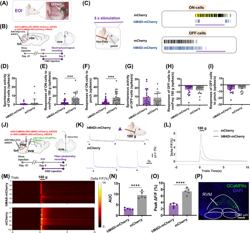
A dental crown produces experimental bite interference and hypersensitivity over one week. Recipient inhibition during model development reduces day-seven RVM ON mechanical and pinch responses and population calcium, without significant spontaneous ON or OFF-cell changes.
Activation in nonmodel rats and inhibition during interference provide complementary evidence. ON-cell selectivity appears in both directions, linking LPBN-recipient activity with medullary stimulus facilitation.
Source: Mo S et al. · The journal of headache and pain · CC BY-NC-ND
Original figure and English caption ↗
Sp5 responses to stronger stimuli also decline
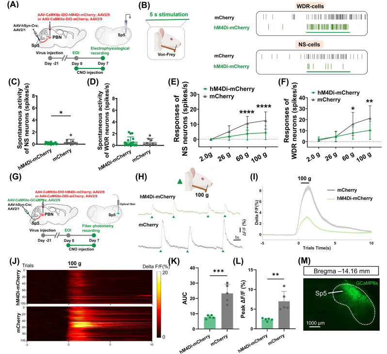
The same arrangement reduces stronger-stimulus WDR/NS responses and Sp5 calcium peaks and areas. Spontaneous activity decreases in NS cells but not significantly in WDR cells. Inhibition selectively attenuates shared stronger-stimulus responses.
Medullary and trigeminal responses both change with LPBN manipulation, indicating coordinated network effects. Physiological recordings are anesthetized, while behavioral testing supplies complementary evidence about model expression.
Source: Mo S et al. · The journal of headache and pain · CC BY-NC-ND
Original figure and English caption ↗
Improved thresholds and preference for inhibition-associated context
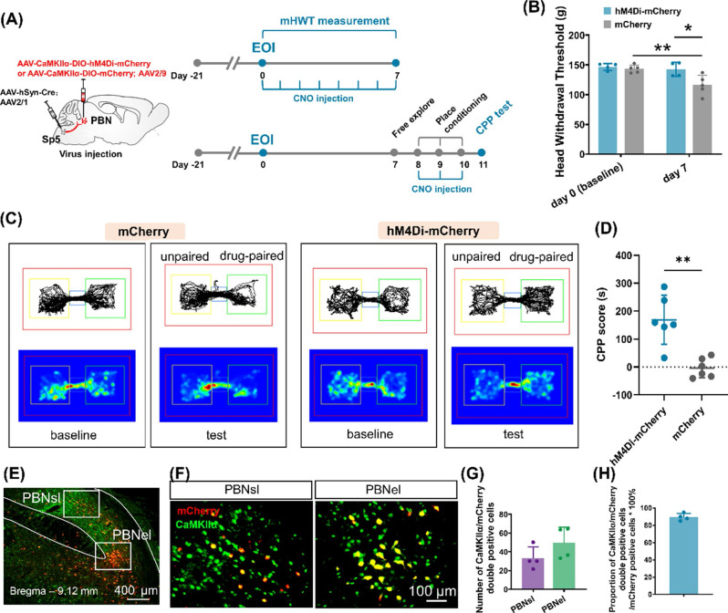
Day-seven mechanical thresholds are higher with repeated inhibition than in model controls. A separate cohort conditioned after hypersensitivity develops prefers the inhibition-associated compartment. These assess intervention during development and place choice in an established hypersensitive state.
The male SD-rat study links bite-related sensitivity and avoidance with central processing: Sp5-input-associated LPBN activity influences RVM and Sp5 stimulus responses. The intervening LPBN transfer remains unresolved, while functional results show interacting brainstem networks modulating incoming orofacial signals.
Source: Mo S et al. · The journal of headache and pain · CC BY-NC-ND
Original figure and English caption ↗