Why does an uninjured hindpaw become sensitive? A cortical microglial clue
After forepaw amputation, mice develop mechanical hypersensitivity in the uninjured ipsilateral hindpaw. Cortical microglial changes link neighboring sensory regions with inhibitory-synapse loss and neuronal hyperactivity.
Why can an uninjured body part become more sensitive after injury elsewhere? A study published online in Cell Reports on February 17, 2024 followed mechanical hypersensitivity in the uninjured ipsilateral hindpaw after forepaw amputation in adult male mice. Beyond neuronal changes, microglial alterations appeared across forelimb and hindlimb sensory cortical regions. In hindlimb cortex, increased microglial association with inhibitory synaptic components accompanied weaker neuronal inhibition, offering a cellular explanation for secondary mechanical hypersensitivity.
Cross-regional microglial changes, CCL2–CCR2 signaling and engulfment of inhibitory synaptic components contribute to secondary hindpaw hypersensitivity, with different intervention effects at residual forepaw and hindpaw sites.
Contents · 7 sections
Forepaw injury precedes a hindpaw threshold change
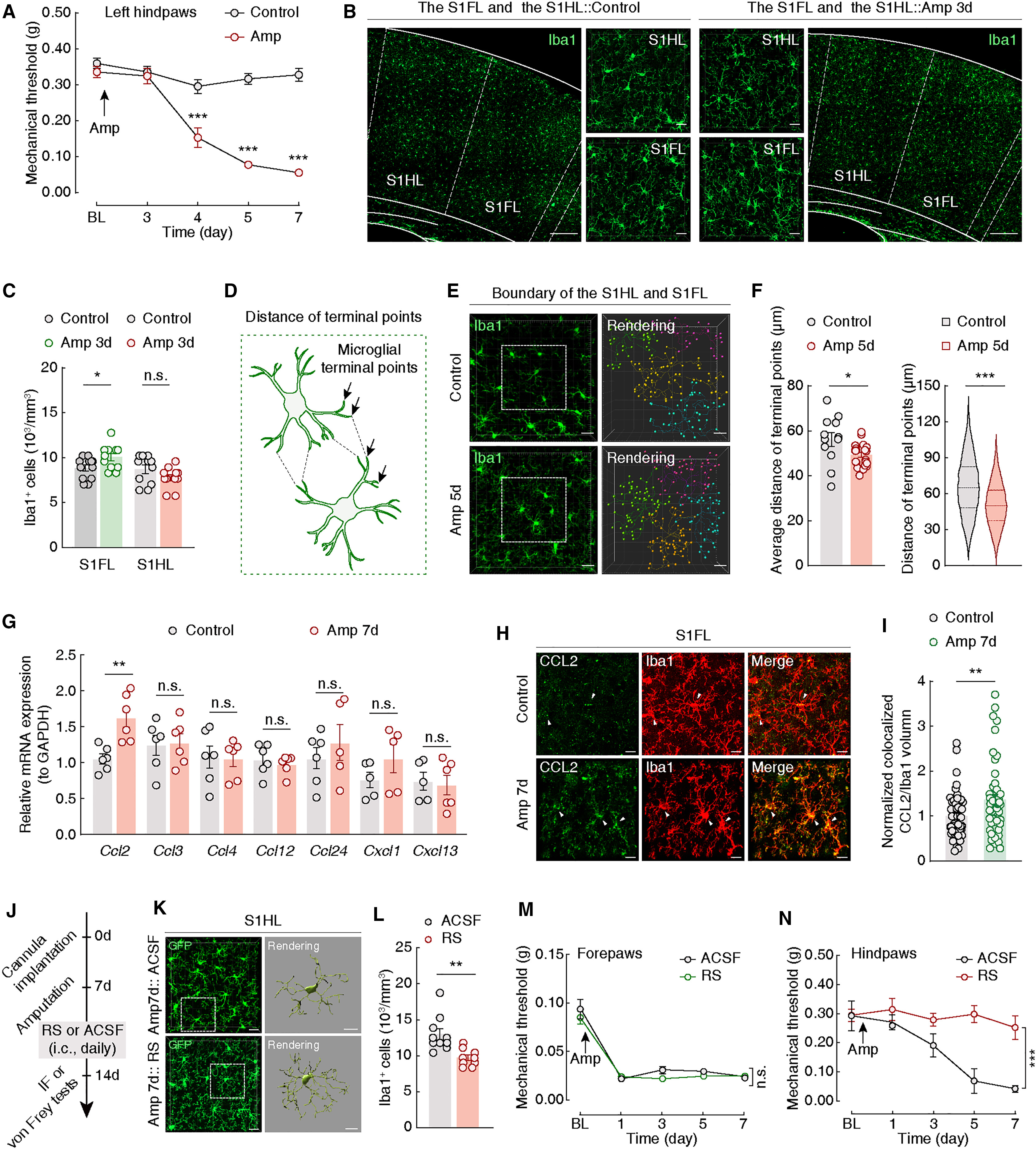
Residual forepaw withdrawal thresholds fell early after amputation, whereas ipsilateral hindpaw changes appeared later and persisted during observation. The uninjured hindpaw therefore showed secondary mechanical sensitization, measured through withdrawal behavior rather than a verbal pain report.
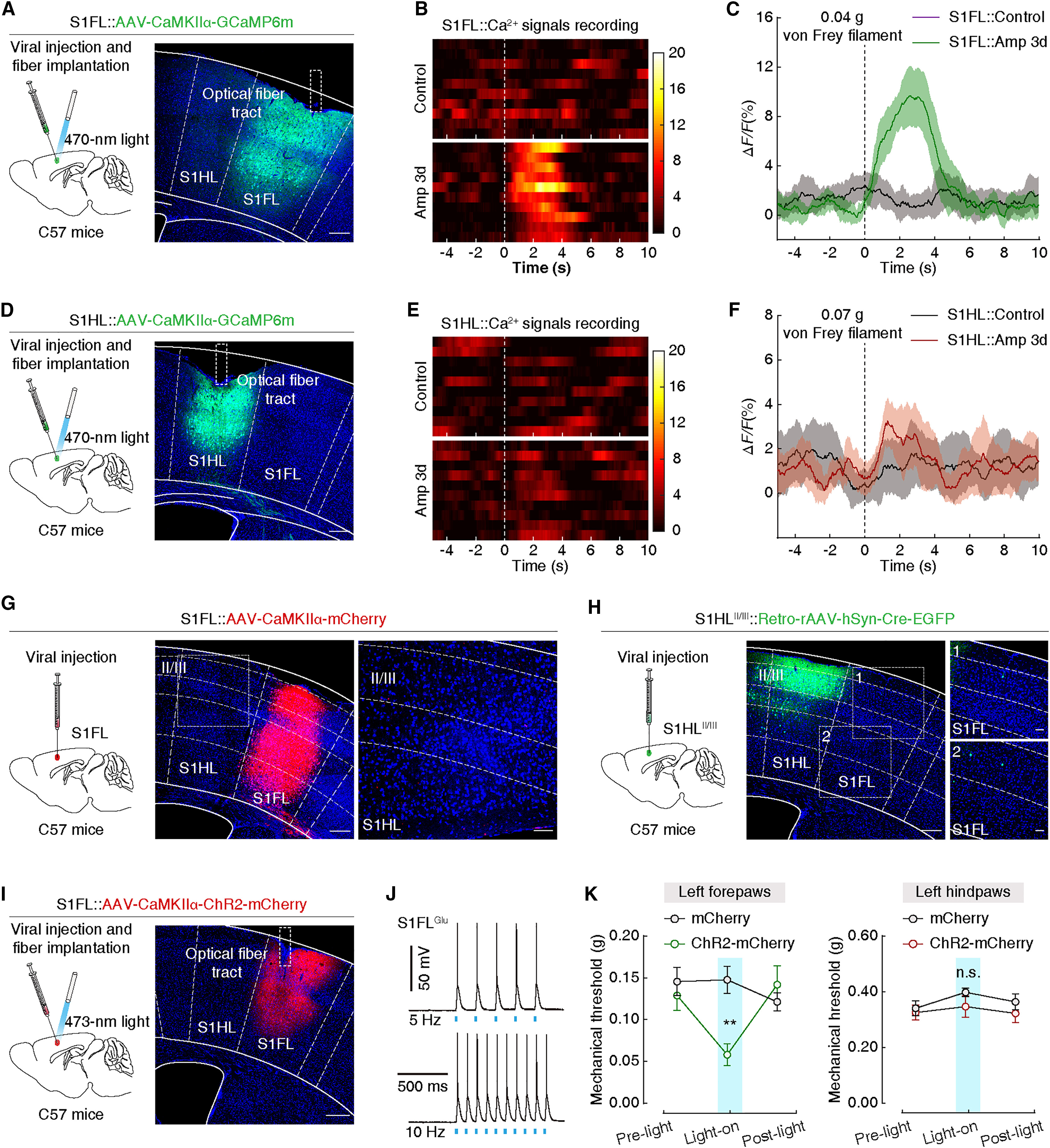
Forelimb and hindlimb information correspond to S1FL and S1HL sensory cortical regions. Excitatory activity was higher in both at the later stage, while earlier changes were concentrated in forelimb cortex. Their timing raised the question of how the regions were linked.
Acute activation does not reproduce the hindpaw change
A direct explanation would be stronger excitation passing from forelimb to hindlimb cortex. The examined pathways did not reveal an obvious S1FL projection to superficial S1HL excitatory neurons. Acute S1FL excitation lowered forepaw thresholds without a significant ipsilateral hindpaw change.
This leaves slower or indirect contributions from forelimb cortex open. It directed attention toward microglia, which maintain the brain environment and interact with synapses. Cross-regional effects may therefore involve processes beyond a direct neuronal projection.
Source: Hong-Rui Wei et al. · Cell Reports · CC BY-NC-ND 4.0; © 2024 The Author(s).
Original figure and English caption ↗
Cellular changes connect with behavior
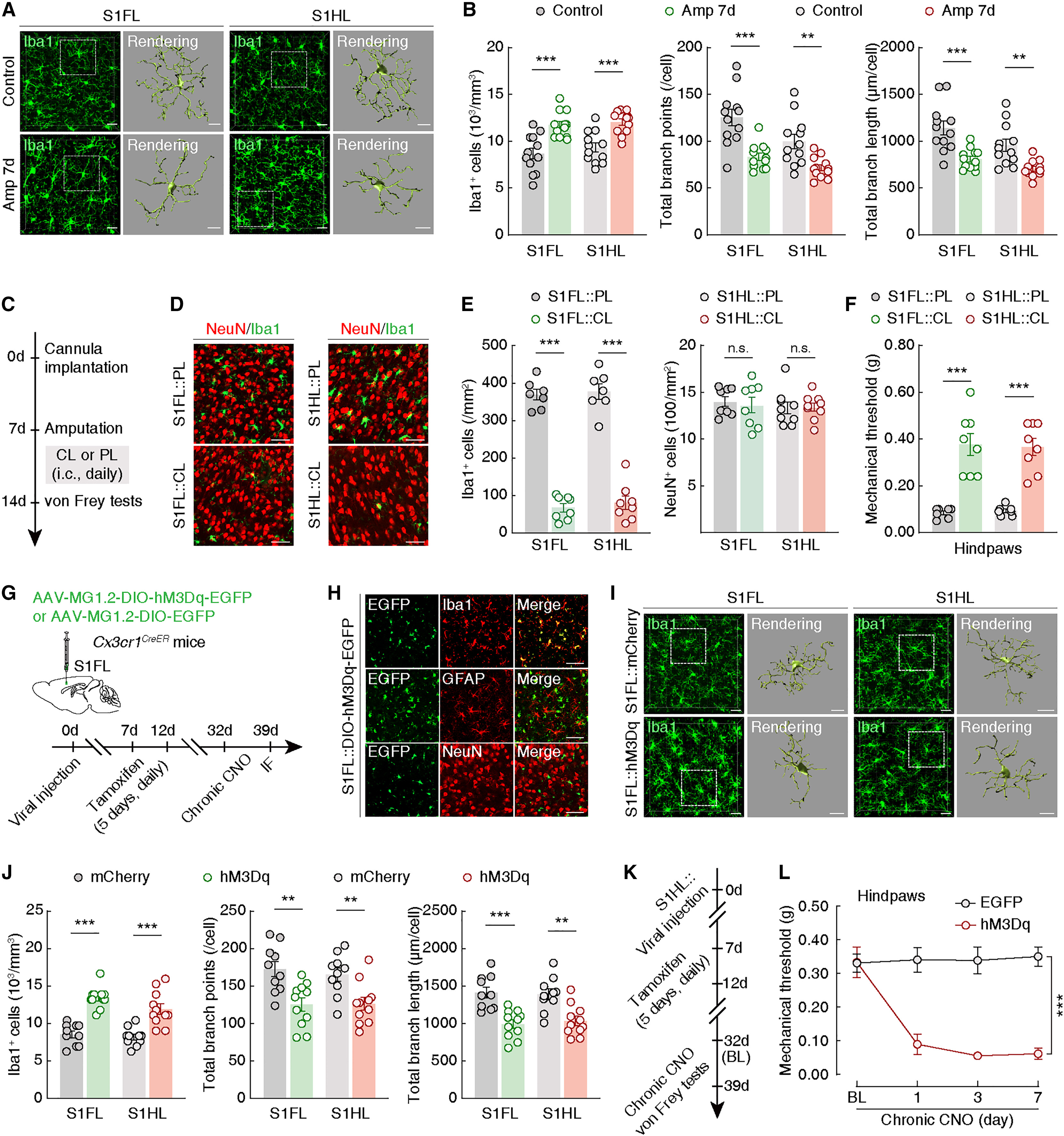
At the secondary-sensitivity stage, both regions showed more microglial labeling, fewer branch points and shorter processes. Local microglial depletion in either region raised ipsilateral hindpaw thresholds without a corresponding significant residual-forepaw effect. Local neuronal-marker counts did not change significantly.
Other functional comparisons linked increased forelimb-region microglial activity with hindlimb-region changes. Directly increasing related activity in hindlimb cortex also lowered hindpaw thresholds. Morphology and functional findings thus implicated microglia in secondary sensitization.
Source: Hong-Rui Wei et al. · Cell Reports · CC BY-NC-ND 4.0; © 2024 The Author(s).
Original figure and English caption ↗
CCL2–CCR2 provides a molecular link
At an early stage, microglial numbers increased in forelimb cortex without a significant corresponding hindlimb-region count change. Later, process tips near the boundary were closer, suggesting altered interaction opportunities rather than directly observed cell migration.
Forelimb cortical tissue showed higher Ccl2 expression and greater CCL2 overlap with microglial labeling. Local CCR2 antagonism reduced hindlimb-region microglial changes and raised hindpaw thresholds, without significant residual-forepaw improvement. This supports a contribution from the pathway while leaving other signals open.
Source: Hong-Rui Wei et al. · Cell Reports · CC BY-NC-ND 4.0; © 2024 The Author(s).
Original figure and English caption ↗
Less inhibitory input reaches excitatory cells
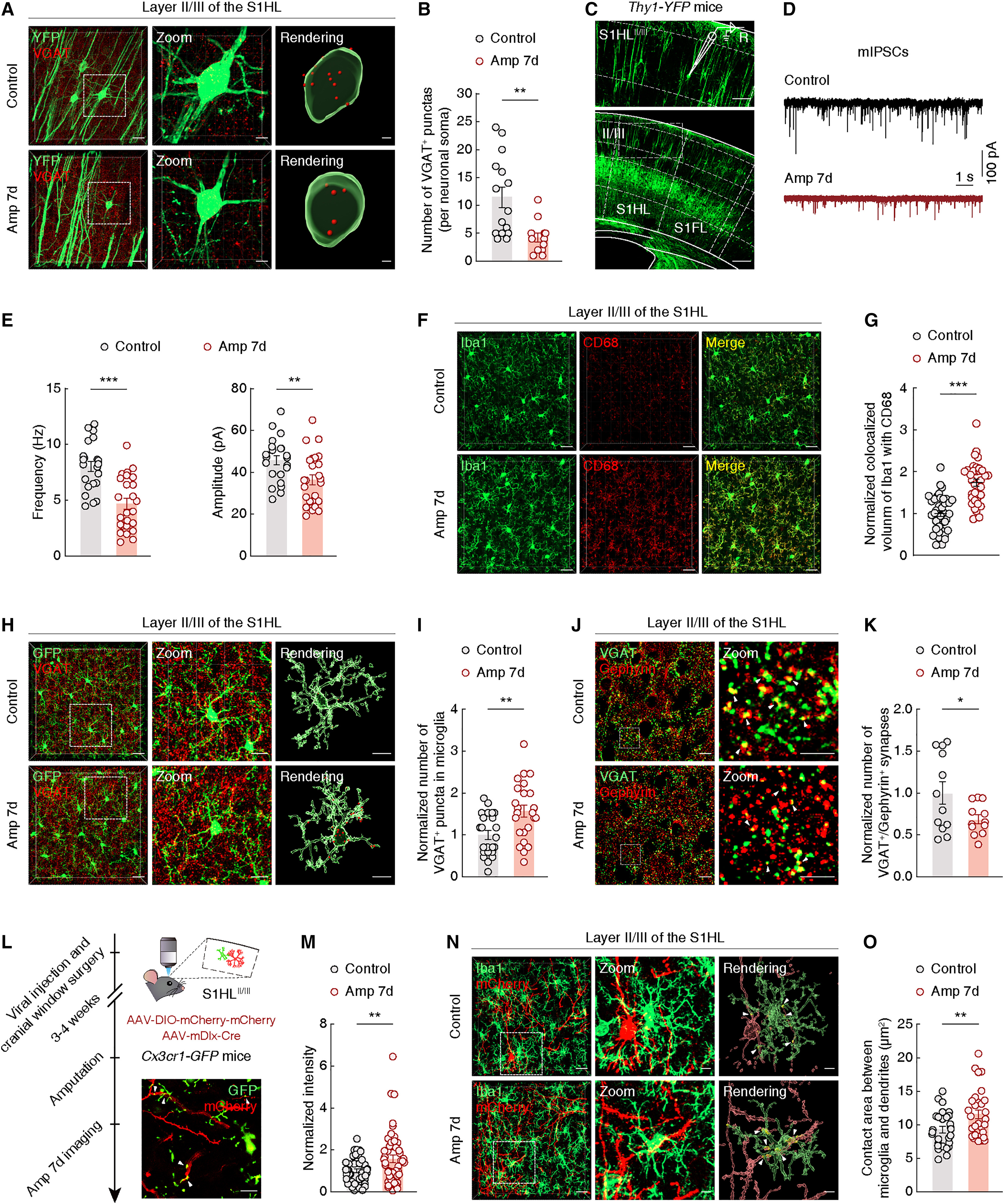
VGAT-labeled inhibitory presynaptic components were reduced around hindlimb cortical excitatory neurons. Miniature inhibitory postsynaptic currents had lower frequency and amplitude, providing convergent structural and electrical evidence of reduced inhibition.
Microglia showed more phagocytosis-associated labeling and greater overlap and engulfment of VGAT components, while paired inhibitory pre- and postsynaptic labeling decreased. The findings implicate enhanced engulfment in disinhibition; why these components are selected remains unresolved.
Source: Hong-Rui Wei et al. · Cell Reports · CC BY-NC-ND 4.0; © 2024 The Author(s).
Original figure and English caption ↗
Hindpaw improvement does not extend to every readout
Under microglia-associated inhibition, stimulus-related calcium responses fell in hindlimb cortical excitatory cells without a significant corresponding forelimb-region change. Inhibitory-current frequency increased, but amplitude did not significantly increase, indicating improvement across selected readouts.
Behavior retained the site difference: ipsilateral hindpaw thresholds rose without a significant residual-forepaw change. This converged with local cellular and receptor-related interventions while separating secondary sensitization from the residual injury-site response.
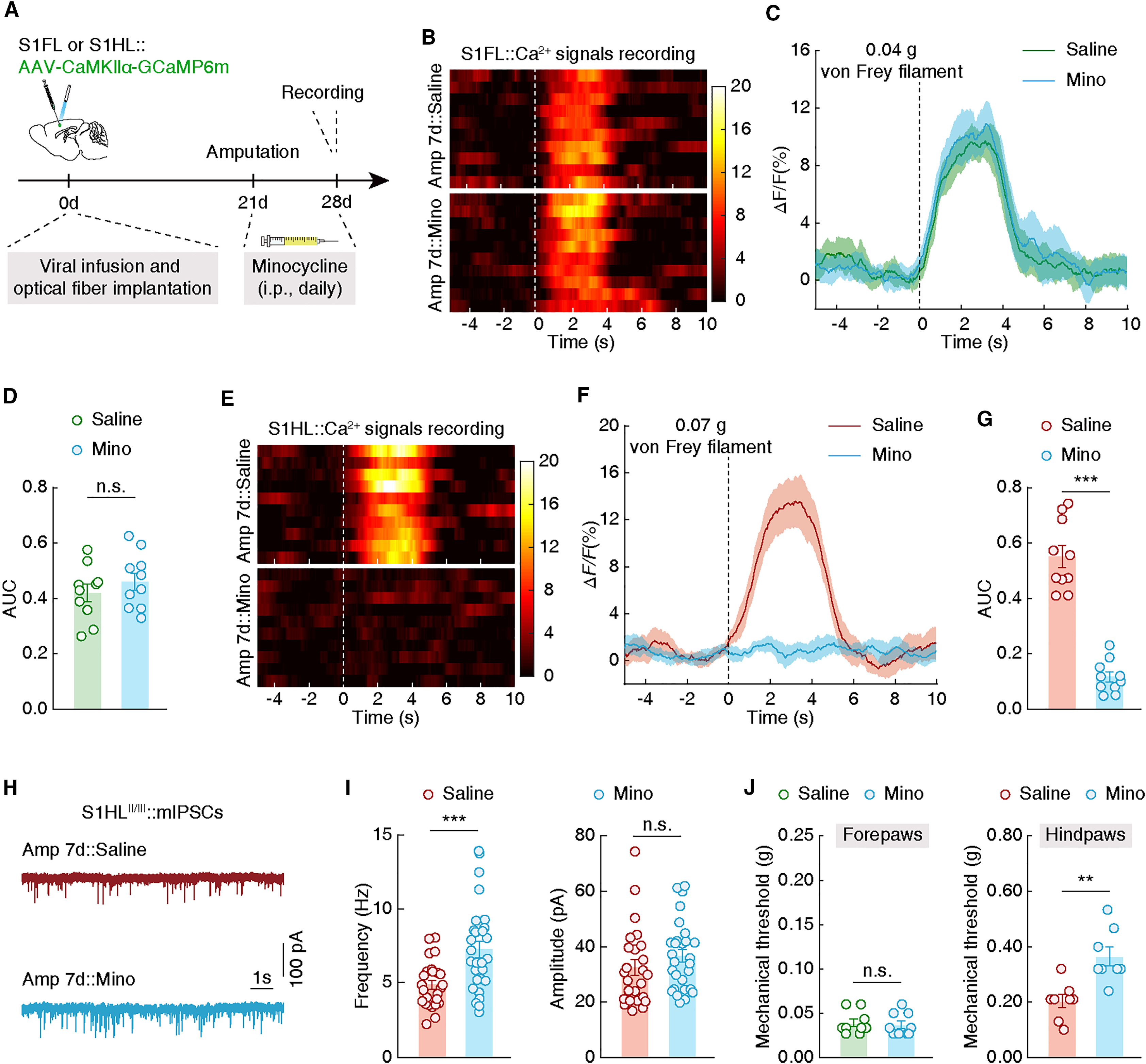
Source: Hong-Rui Wei et al. · Cell Reports · CC BY-NC-ND 4.0; © 2024 The Author(s).
Original figure and English caption ↗
Microglia add a layer to cross-regional interaction
ACC-directed hindlimb cortical output contributed to hindpaw sensitivity, with partial threshold improvement after inhibition. Inhibiting terminals in lPAG did not significantly change either paw threshold. Distinct outputs therefore contribute differently within the broader network.
The study concerns secondary mechanical hypersensitivity in an uninjured mouse hindpaw, which differs from subjective human phantom pain. It provides a cortical cellular explanation for sensitivity beyond an injury site and raises questions about signals controlling the process and selectively limiting maladaptive changes.