Hypothalamic cells participate in abdominal sensitivity in tumor-bearing mice
In pancreatic-tumor mice, labeled excitatory PVN cells are more excitable. Reducing their number or activity weakens abdominal mechanical sensitivity at selected times, with different effects on hunching and later outcomes.
Pain associated with pancreatic tumors raises a question beyond local tissue: how does the brain participate? A 2024 Hepatobiliary Surgery and Nutrition study examines hypothalamic paraventricular nucleus, PVN, in male mice. This region connects autonomic and stress regulation and contains diverse cells. Following abdominal behavior, tracing and activity measurements, the team finds increased excitability in a labeled excitatory population. Reducing its number or activity weakens selected abdominal responses, with effects varying across measures and observation times.
In a pancreatic-tumor model, labeled excitatory PVN cells fire more and show population calcium responses to abdominal stimulation. Reducing or suppressing the population attenuates mechanical sensitivity at selected observations; final mechanical and hunch comparisons show no significant improvement.
Contents · 8 sections
Abdominal sensitivity emerges before hunching changes
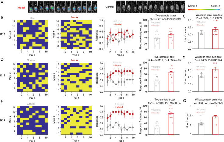
Tumor-model mice respond more frequently to abdominal mechanical stimulation. At an early assessment, sensitivity differs from controls while hunch scores do not significantly differ. Both measures change at later observations, with wider group differences.
Mechanical responses and posture describe discomfort-related behavior from different angles. Their timing differs, and the study follows this progression to identify central cells participating in abdominal responses.
Source: Ji NN et al. · Hepatobiliary surgery and nutrition · CC BY-NC-ND
Original figure and English caption ↗
Tracing identifies PVN within a multisynaptic network
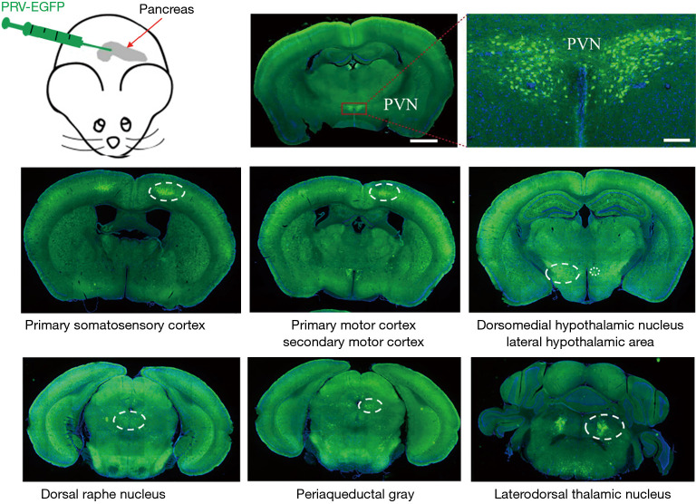
Retrograde transsynaptic tracing from pancreas labels PVN cells and several cortical and brainstem regions. PVN emerges within a candidate network, connected to pancreas through multiple stages rather than one direct link.
PVN participates in autonomic and stress-related regulation, linking bodily state with central activity. The study next compares activity labeling across PVN cells and records their excitability and responses during abdominal stimulation.
Source: Ji NN et al. · Hepatobiliary surgery and nutrition · CC BY-NC-ND
Original figure and English caption ↗
Activity labeling directs attention to excitatory cells
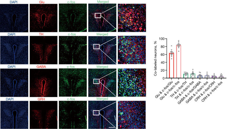
PVN contains multiple cell populations. Comparing c-Fos with cellular markers shows greater overlap with glutamatergic labeling than with other tested markers, directing further study toward an excitatory population.
Colabeling describes activity associations within sampled cells. Glutamatergic cells are already abundant in PVN, and transmitter and peptide markers may overlap. The finding identifies a population to investigate without fully separating all cellular identities.
Source: Ji NN et al. · Hepatobiliary surgery and nutrition · CC BY-NC-ND
Original figure and English caption ↗
Matched current elicits more firing
Slice recordings show higher firing with matched current in labeled excitatory PVN cells from model mice, together with a more depolarized resting membrane potential. Their responses to input have changed.
Action-potential thresholds do not significantly differ. Rather than every electrophysiological measure increasing, a specific pattern of excitability changes complements the activity labeling and clarifies cellular state.
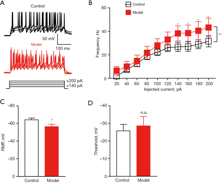
Source: Ji NN et al. · Hepatobiliary surgery and nutrition · CC BY-NC-ND
Original figure and English caption ↗
Population calcium rises around abdominal stimulation
Abdominal stimulation in tumor-model mice accompanies increased calcium in the labeled excitatory PVN population, unlike the control fluorophore. Mean and peak comparisons support an event-related change, linking population activity with the tested stimulation in time.
These recordings compare a responsive calcium indicator with a control fluorophore, describing population changes around stimulation. They complement slice excitability measurements: one follows event-related activity, while the other characterizes responses to input current.
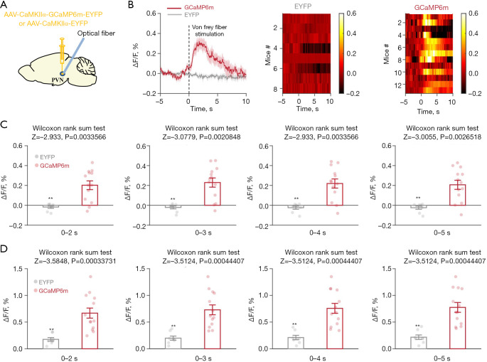
Source: Ji NN et al. · Hepatobiliary surgery and nutrition · CC BY-NC-ND
Original figure and English caption ↗
Reducing cells attenuates sensitivity at selected times
After reducing labeled excitatory PVN cells, abdominal mechanical responses decrease at the two earlier assessments. Hunch scores do not significantly change at either time; at the final assessment, neither measure significantly differs between groups.
The two discomfort-related measures respond differently: mechanical responses weaken earlier, while hunching shows no corresponding change. The relationship between this population and bodily behavior therefore varies across measured outcomes.
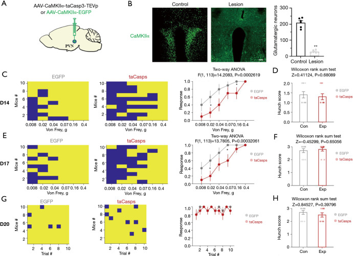
Source: Ji NN et al. · Hepatobiliary surgery and nutrition · CC BY-NC-ND
Original figure and English caption ↗
Suppressing activity has measure- and time-dependent effects
A separate experiment suppresses population activity, again reducing mechanical responses at the two earlier assessments. Hunch scores decrease only at the second of those times. Neither final measure significantly improves, giving a pattern related to but distinct from cell reduction.
As tumors progress, overall animal condition may also affect later responses. This stage difference highlights the need to relate later sensitivity to overall condition. Mechanical responses and hunching do not always change together, making the link between cellular activity and specific behavior more detailed than one overall score.
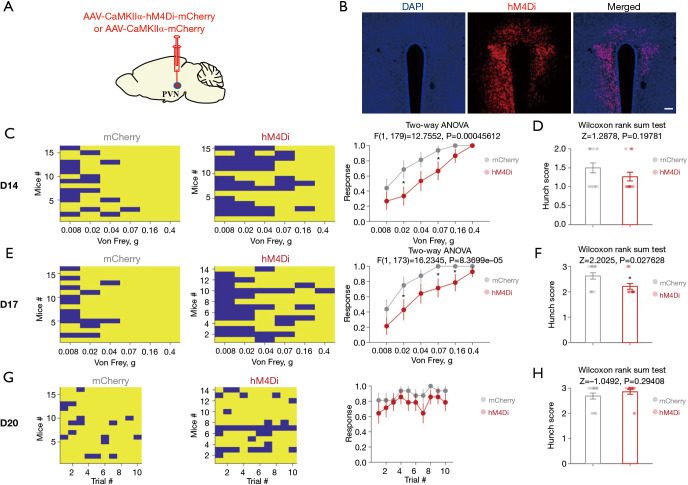
Source: Ji NN et al. · Hepatobiliary surgery and nutrition · CC BY-NC-ND
Original figure and English caption ↗
Multiple inputs converge on a bodily-regulation node
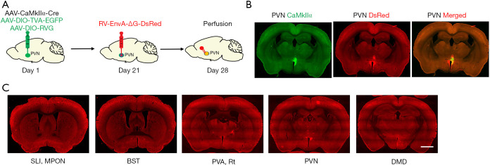
Tracing backward from labeled excitatory PVN cells identifies inputs from preoptic, bed-nucleus, thalamic and hypothalamic regions. These form a structural network around the population, without individually establishing which input drives the tested abdominal responses.
This male-mouse study connects pancreatic-tumor-related behavior with hypothalamic cells. Central changes in visceral discomfort concern regional networks and the excitability of specific populations. Differences across stages and measures give that connection a more precise shape.
Source: Ji NN et al. · Hepatobiliary surgery and nutrition · CC BY-NC-ND
Original figure and English caption ↗