Why do ventral pallidal cells respond differently when acetylcholine rises?
A mouse study follows STN input into cell-specific receptor changes during chronic hypersensitivity, revealing an intervention with different effects before and after injury.
When one transmitter rises, do its recipient cells become more active together? In the injured mouse ventral pallidum, cholinergic and GABAergic cells become more excitable while glutamatergic cells become less so. Researchers followed STN input, acetylcholine and nicotinic receptors to understand the divergence.
After nerve injury, acetylcholine signals rise and receptor changes differ by cell type. Reducing GABAergic-cell α4 limits severe hypersensitivity but lowers baseline thresholds before injury.
Contents · 6 sections
A motor-associated network also regulates noxious responses
The subthalamic nucleus, often studied in motor control, also participates in responses to noxious stimuli. The ventral pallidum contains cholinergic, GABAergic and glutamatergic cells with different local and distant outputs. Earlier stimulation findings did not establish whether STN and VP cholinergic neurons formed one functional chain.
The questions became specific: does STN influence withdrawal through VP cholinergic cells, and how do the connection and local transmitter change after injury? Following input, release, cells and receptor subunits revealed diverging responses across populations.
How does STN input recruit VP cholinergic cells?
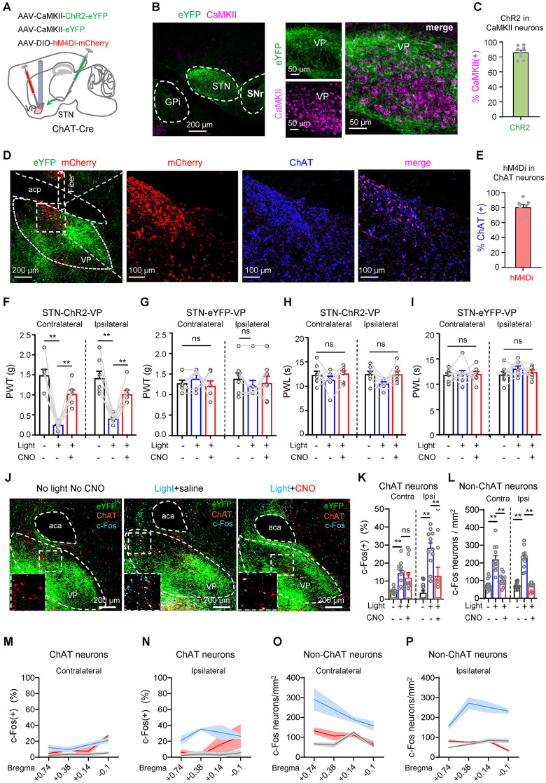
Tracing identified STN as one input to VP cholinergic cells. Terminal stimulation evoked excitatory currents with pharmacology supporting a monosynaptic glutamatergic connection. Unilateral STN–VP stimulation lowered mechanical withdrawal thresholds on both hind paws, and VP cholinergic inhibition attenuated the behavioral and local activity effects.
Unilateral STN–VP stimulation altered mechanical thresholds without significantly shortening thermal withdrawal latency. Direct activation of STN-recipient VP cholinergic cells produced both mechanical and thermal sensitization, blocked by local nicotinic antagonism. Input and recipient activation had related but distinct effects.
Source: Ji YW et al. · PLoS biology · CC BY
Original figure and English caption ↗
More transmitter, divergent cellular responses
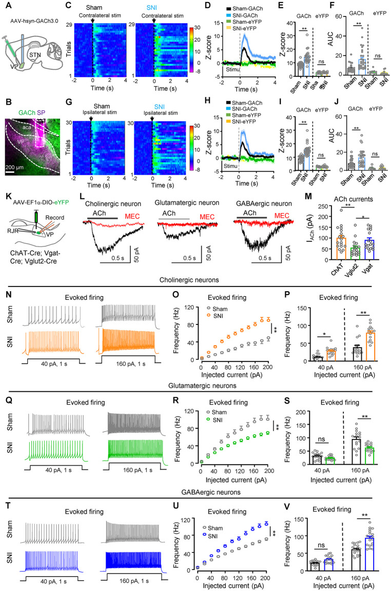
In spared nerve injury mice, mechanical stimulation evoked stronger VP cholinergic calcium responses, reduced by STN or VP-projecting STN inhibition. Acetylcholine-sensor responses also increased after injury and were attenuated by local cholinergic inhibition. Voluntary paw movement controls helped separate sensory stimulation from accompanying movement.
Slices showed increased cholinergic and GABAergic excitability but decreased glutamatergic excitability. STN inhibition weakened some injury-related responses while differences in decay and area remained. Opposite cellular states alongside an increased acetylcholine signal made receptor localization the next focus.
Source: Ji YW et al. · PLoS biology · CC BY
Original figure and English caption ↗
Receptor changes depend on the recipient cell
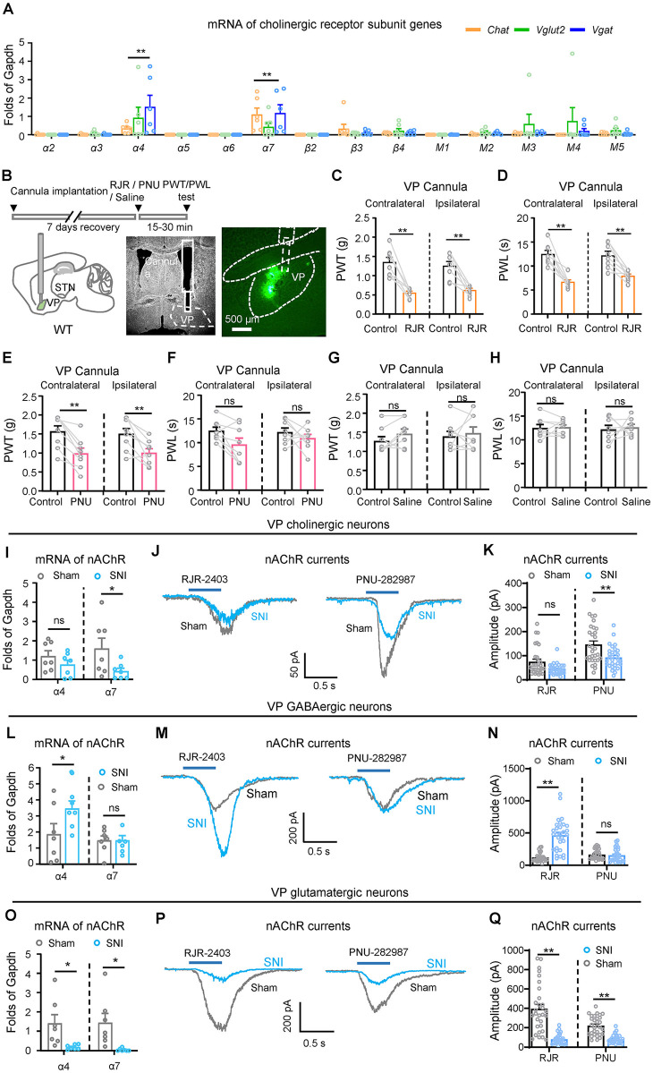
After injury, α4-containing nicotinic receptors increased in GABAergic cells but decreased in glutamatergic cells. α7-containing receptors decreased in cholinergic and glutamatergic cells. Cell-resolved gene measurements and evoked currents revealed distinct receptor trajectories.
In normal mice, local α4 or α7 blockade left baseline mechanical and thermal thresholds unchanged. α4 activation lowered both, while α7 activation mainly lowered mechanical thresholds, leaving thermal latency comparable. Baseline and induced states therefore revealed different receptor effects.
Source: Ji YW et al. · PLoS biology · CC BY
Original figure and English caption ↗
An unexpected combination: baseline sensitization and reduced injury effects
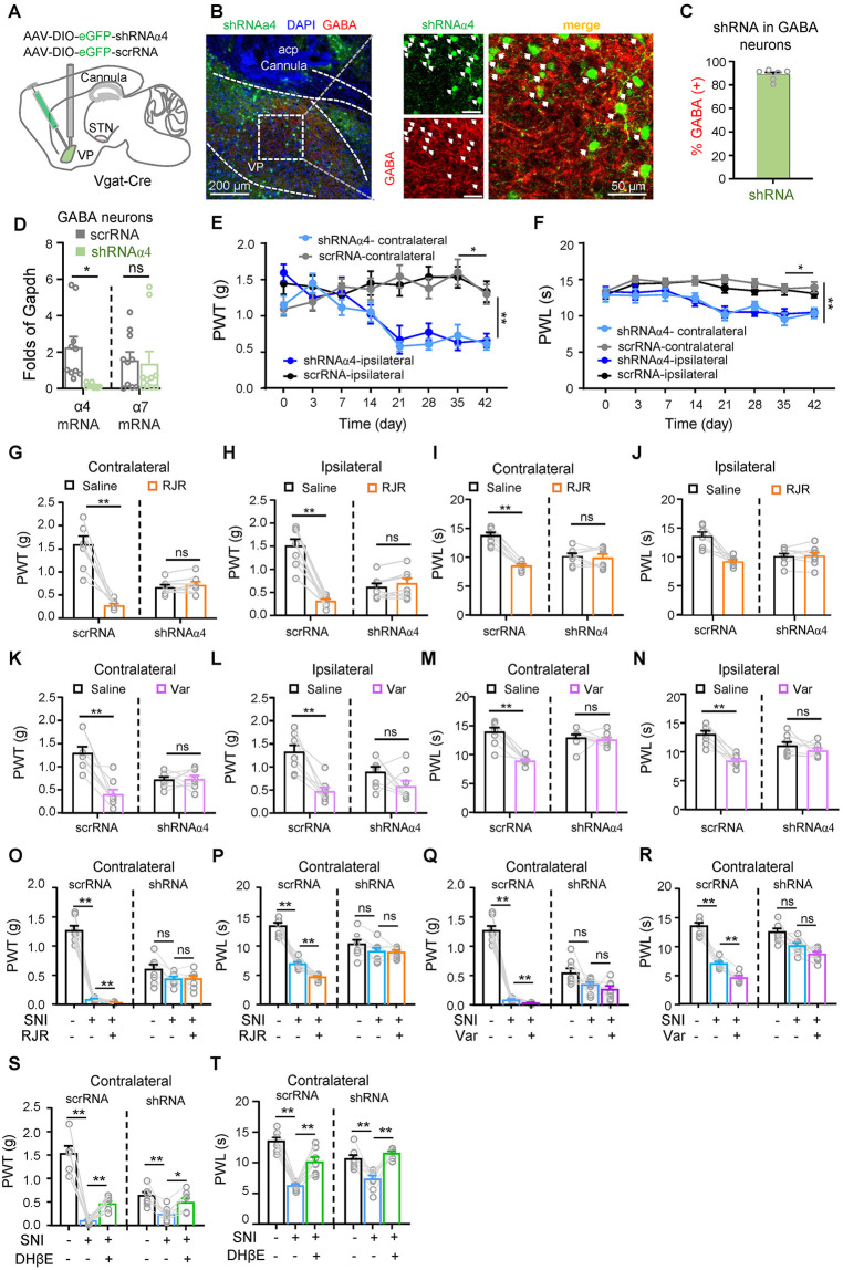
Reducing GABAergic α4 blocked further agonist-induced threshold reductions and limited severe sensitization after SNI. Yet before injury, the same manipulation modestly lowered mechanical and thermal thresholds. One receptor change affected both baseline responsiveness and the magnitude of injury effects.
Increasing glutamatergic-cell α4 or α7 did not improve post-injury baseline hypersensitivity, but enhanced threshold-raising responses to the corresponding antagonists; naloxone did not block these effects. Cell location and network state jointly shaped receptor influence.
Source: Ji YW et al. · PLoS biology · CC BY
Original figure and English caption ↗
Reading hypersensitivity through cells and state
Supplementary tracing identified cross-hemisphere non-cholinergic VP connections relevant to bilateral withdrawal effects. STN input, local acetylcholine and cell-specific receptors form a network in which one transmitter can lead to different cellular states.
The study primarily used male mice with SNI. Full receptor composition, adaptation during prolonged manipulation and the contrasting baseline/injury effects remain open. These questions will guide attempts to modulate hypersensitivity while preserving normal responses.