Why do sounds become aversive after nerve injury? A cross-modal link between sensory cortices
A mouse study finds that peripheral nerve injury strengthens direct excitatory input from hindlimb somatosensory cortex to auditory cortex, driving sound hypersensitivity.
Patients with chronic neuropathic pain frequently experience heightened sensitivity to everyday environmental sounds (hyperacusis) alongside somatosensory allodynia, yet how peripheral nerve injury cross-modally reshapes central auditory processing has remained unclear. On January 30, 2025, Zhi Zhang, Haitao Wang, Yuanyin Wang, and colleagues at the University of Science and Technology of China and Anhui Medical University published a research article in Current Biology entitled 'Cross-modal cortical circuit for sound sensitivity in neuropathic pain' (with Yunfeng Mao and Mingjun Zhang as co-first authors). Combining head-fixed pupillometry, Go/No-Go auditory discrimination tasks, in vivo fiber photometry, multi-channel silicon probe recordings, viral circuit tracing, and opto-/chemogenetic manipulations, the team identified a direct monosynaptic projection from hindlimb primary somatosensory cortex glutamatergic neurons to auditory cortex glutamatergic neurons (S1HLGlu→ACxGlu) that drives sound hypersensitivity under neuropathic pain.
Key finding: Seven days after spared nerve injury (SNI), mice exhibit both mechanical allodynia and heightened sound sensitivity—manifested as amplified noise-evoked pupil dilation and shortened lick reaction latency in Go/No-Go tasks across 40–60 dB SPL. SNI-induced hyperactivity of hindlimb primary somatosensory cortex glutamatergic neurons (S1HLGlu) directly excites auditory cortex glutamatergic neurons (ACxGlu) via a monosynaptic pathway, and bidirectional chemogenetic manipulation of this S1HLGlu→ACx circuit regulates sound sensitivity in naive and SNI mice.
Contents · 5 sections
Peripheral neuropathic pain induces amplified sound-evoked pupil dilation and shortened auditory behavioral latency in mice
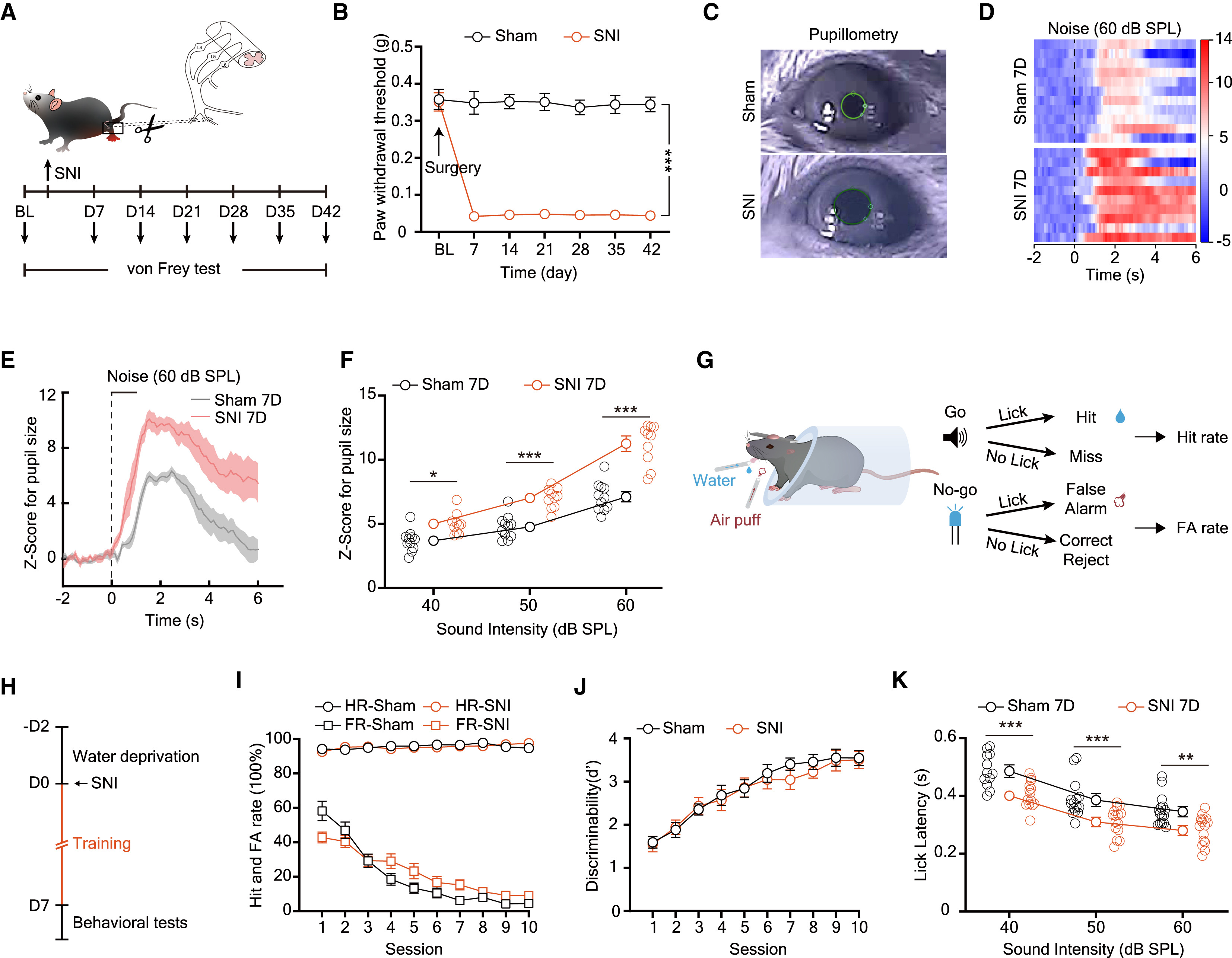
To quantitatively assess whether peripheral nerve injury alters auditory sensitivity, the authors established the spared nerve injury (SNI) model in mice, which produced robust mechanical allodynia from day 7 through day 42 post-surgery. At 7 days post-SNI (SNI 7D), infrared pupillometry in head-fixed awake mice revealed that 40, 50, and 60 dB SPL white noise bursts evoked significantly larger pupil dilation Z-scores in SNI 7D mice than in Sham 7D controls, demonstrating heightened autonomic and sensory reactivity to innocuous sound stimuli.
Using a water-restricted Go/No-Go auditory discrimination task (auditory cue as the Go water-reward signal and blue light flash as the No-Go air-puff signal), the authors found that SNI and Sham mice achieved comparable hit rates, false alarm rates, and discriminability indices (d') across 10 training sessions. However, during post-SNI day 7 testing across 40, 50, and 60 dB SPL sound intensities, SNI 7D mice exhibited significantly shorter lick latencies following tone onset than Sham controls, confirming behavioral hyperacusis.
Source: Yunfeng Mao et al. · Current Biology · © Elsevier Inc.
Original figure and English caption ↗
In vivo calcium imaging and silicon probe recordings reveal selective hyperexcitability of auditory cortex glutamatergic neurons after SNI
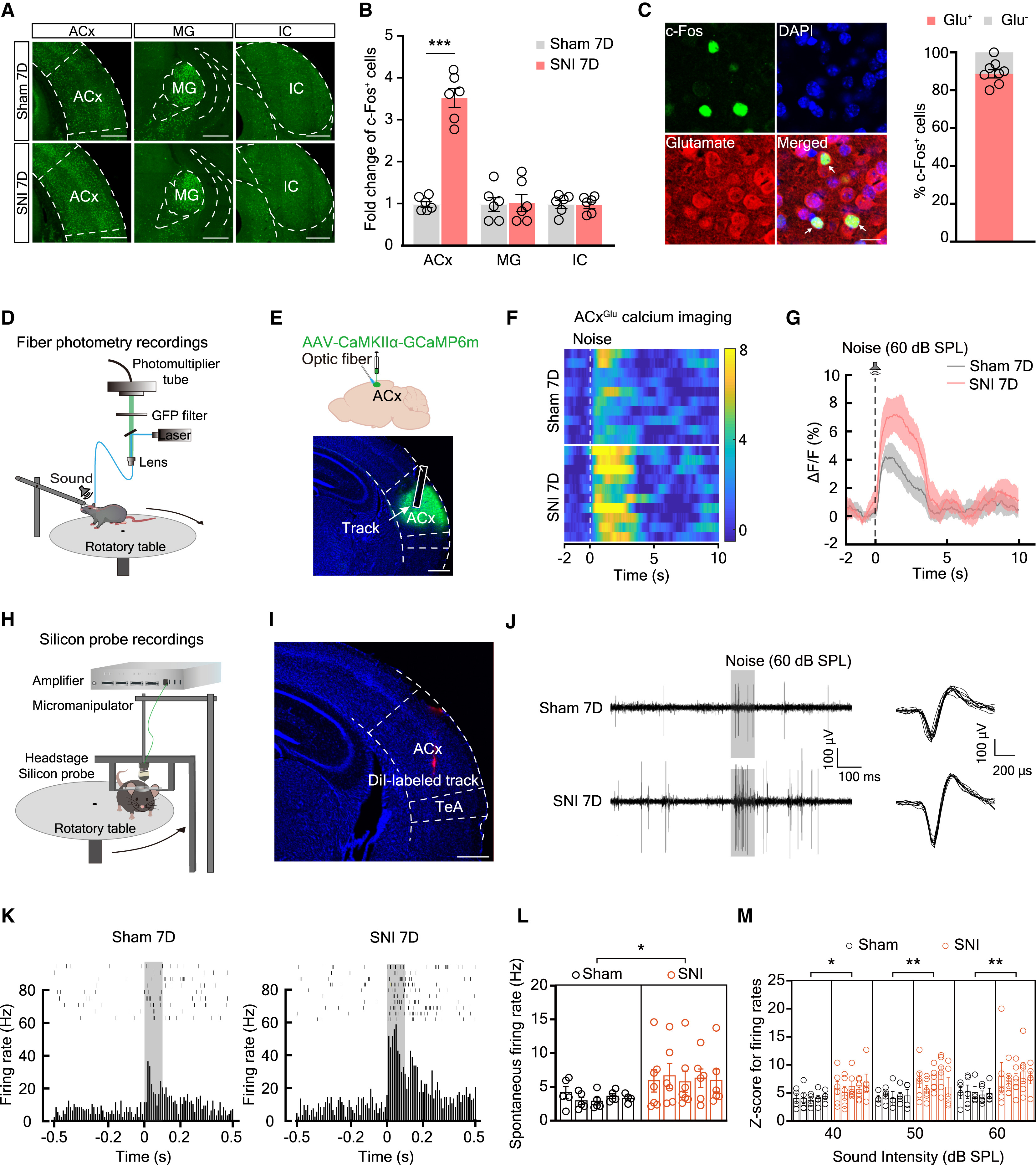
To pinpoint the central locus of pain-induced auditory hypersensitivity, the authors mapped noise-evoked c-Fos expression across the auditory pathway. Compared with Sham 7D mice, SNI 7D mice displayed an approximately 3.5-fold increase in c-Fos+ cells selectively within the auditory cortex (ACx), with no significant change in the medial geniculate body (MG) or inferior colliculus (IC); ~90% of ACx c-Fos+ neurons co-expressed glutamate (Glu+). In vivo fiber photometry in mice expressing AAV-CaMKIIα-GCaMP6m in the ACx confirmed significantly elevated noise-evoked calcium transients (ΔF/F) in ACxGlu neurons at SNI 7D.
In vivo multi-channel silicon probe recordings further demonstrated that ACx neurons in SNI 7D mice exhibited significantly higher spontaneous firing rates as well as elevated noise-evoked firing Z-scores across 40, 50, and 60 dB SPL, establishing both spontaneous and sound-evoked hyperexcitability of ACxGlu neurons under neuropathic pain.
Source: Yunfeng Mao et al. · Current Biology · © Elsevier Inc.
Original figure and English caption ↗
Viral tracing and slice optogenetics establish a direct monosynaptic excitatory S1HLGlu→ACxGlu cortical pathway
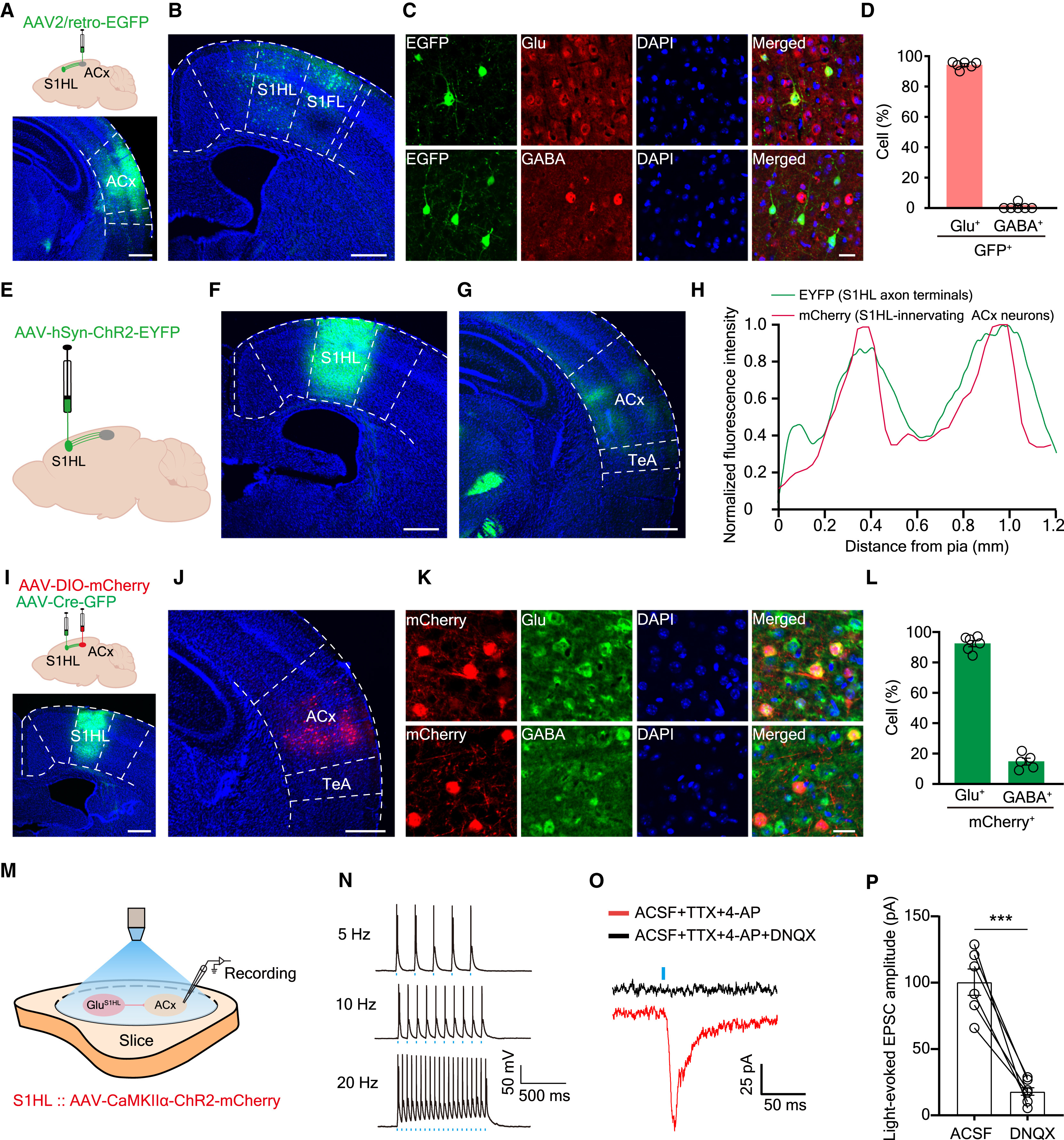
Given the absence of early c-Fos changes in the MG or IC, the authors hypothesized that ACx hyperexcitability arises from direct cross-modal cortical input. Retrograde tracing from the ACx with AAV2/retro-EGFP labeled abundant somatosensory cortical neurons in the hindlimb (S1HL) and forelimb (S1FL) regions, ~94% of which were glutamatergic (Glu+). Anterograde tracing (AAV-hSyn-ChR2-EYFP in S1HL) and anterograde transsynaptic labeling (AAV-Cre-GFP in S1HL + AAV-DIO-mCherry in ACx) confirmed dense S1HL terminal innervation in the ACx targeting predominantly (~92%) Glu+ postsynaptic neurons.
In acute brain slices from mice expressing AAV-CaMKIIα-ChR2-mCherry in the S1HL, blue-light photostimulation (5, 10, 20 Hz) of S1HL axon terminals in the ACx reliably drove action potentials in ACx neurons. In the presence of TTX and 4-AP, photostimulation evoked robust monosynaptic excitatory postsynaptic currents (EPSCs) that were abolished by the AMPA receptor antagonist DNQX, confirming a direct monosynaptic glutamatergic S1HLGlu→ACxGlu synapse.
Source: Yunfeng Mao et al. · Current Biology · © Elsevier Inc.
Original figure and English caption ↗
SNI hyperactivates ACx-projecting S1HL neurons, and activating the S1HLGlu→ACx pathway recapitulates ACx sound hypersensitivity
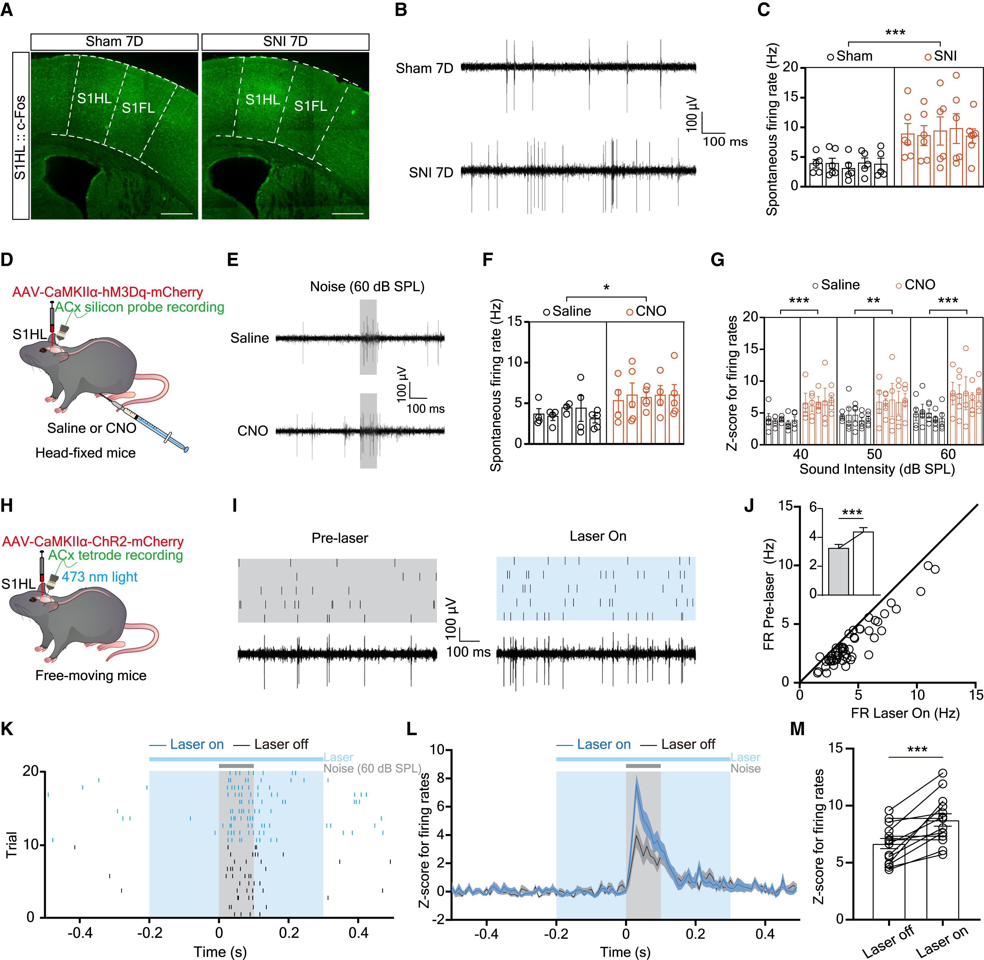
Immunofluorescence co-labeling of AAV2/retro-EGFP-traced ACx-projecting S1HL neurons and in vivo multi-channel recordings revealed significantly increased c-Fos co-localization and elevated spontaneous spike firing rates in the S1HL of SNI 7D mice compared with Sham controls (Figures 4A–C). Chemogenetic activation of S1HLGlu neurons via AAV-CaMKIIα-hM3Dq and systemic CNO (1 mg/kg) in naive mice significantly elevated both spontaneous and noise-evoked firing rates in the ACx during head-fixed multi-electrode recordings (Figures 4D–G).
Furthermore, optrode recordings in freely moving mice expressing AAV-CaMKIIα-ChR2 in the S1HL showed that selective optical stimulation of S1HLGlu axon terminals within the ACx elevated spontaneous ACx firing rates and enhanced noise-evoked firing Z-scores across 40, 50, and 60 dB SPL, demonstrating that S1HLGlu→ACx terminal activation directly amplifies auditory cortical gain.
Source: Yunfeng Mao et al. · Current Biology · © Elsevier Inc.
Original figure and English caption ↗
Bidirectional manipulation of the S1HLGlu→ACx circuit controls behavioral sound sensitivity in naive and SNI mice
At the behavioral level, selective terminal activation via intra-ACx CNO infusion in naive mice expressing AAV-CaMKIIα-hM3Dq in the S1HL significantly increased noise-evoked pupil dilation and shortened lick reaction latencies across 40–60 dB SPL in Go/No-Go tasks compared with saline infusion (Original Figure 5A–D).
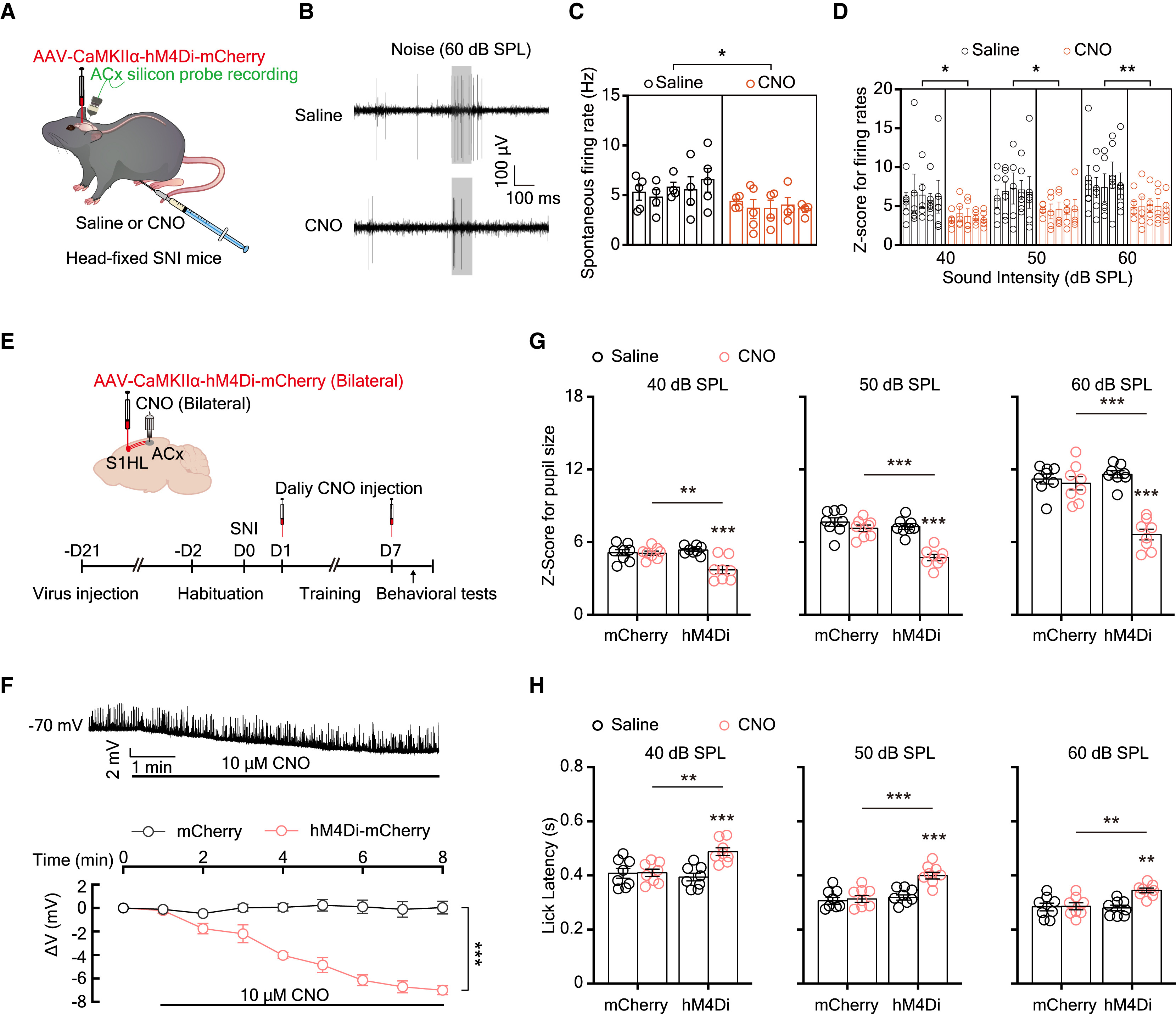
Conversely, chemogenetic inhibition of the S1HLGlu→ACx pathway via AAV-CaMKIIα-hM4Di and intra-ACx CNO infusion in SNI 7D mice (Figures 6A–H) significantly suppressed spontaneous and noise-evoked ACx neuronal hyperactivity, attenuated amplified pupil dilation, and normalized lick reaction latencies. Together, these findings identify a direct monosynaptic somatosensory-to-auditory cortical pathway (S1HLGlu→ACxGlu) that drives sound hypersensitivity during neuropathic pain.
Source: Yunfeng Mao et al. · Current Biology · © Elsevier Inc.
Original figure and English caption ↗