A pain-facilitating cortical connection: greater sensitivity without more anxiety
An excitatory mouse retrosplenial-to-anterior-cingulate connection enhances mechanical and some thermal responses without accompanying changes in emotion-related behavior.
Nociception is more than a straight line carrying stimulation from the body to the brain. Communication between cortical areas can also alter responses. A 2025 PLOS Biology study identifies an excitatory connection from the retrosplenial cortex (RSC) to anterior cingulate cortex (ACC). RSC is often studied in navigation and memory, while ACC participates in nociceptive and emotional processing. Activating their connection increases mouse sensitivity to mechanical and some thermal stimuli without clearly increasing anxiety-like behavior or place aversion.
RSC supplies direct excitatory input to ACC, mediated predominantly by AMPA receptors. Activation enhances selected nociceptive responses; tail-flick after terminal activation and tested emotional behaviors after projection-defined cell activation show no clear changes.
Contents · 7 sections
Who else talks to anterior cingulate cortex?
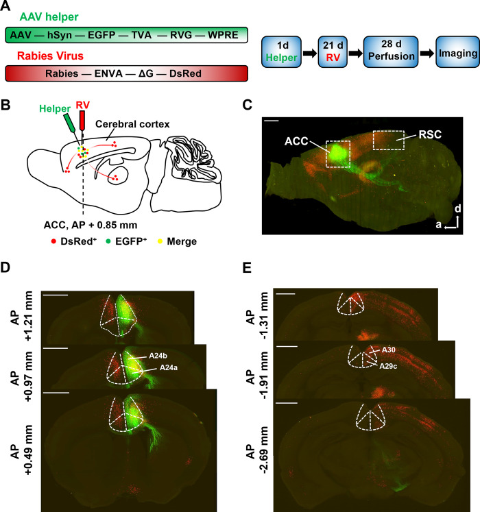
ACC receives input from multiple regions and integrates sensory, emotional and cognitive information. Understanding its role in nociception therefore requires examining who sends signals to it. Whole-brain three-dimensional imaging and retrograde tracing identify RSC among its upstream sources.
Both granular and agranular RSC contain cells projecting to ACC. They occur bilaterally, with more on the same side. Two cortical areas often studied under different themes are thus connected by a route that can be examined functionally.
Source: Hao S et al. · PLoS biology · CC BY
Original figure and English caption ↗
Upstream activation recruits downstream responses
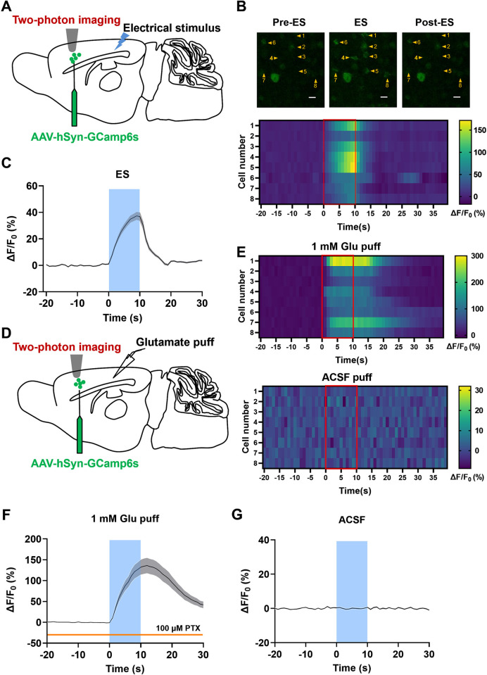
Does a visible connection actually transmit excitation? In slices retaining both regions, RSC stimulation evokes excitatory field responses at multiple ACC recording sites. Calcium imaging independently shows ACC neuronal calcium increasing during RSC activation.
Local activation of RSC cells likewise produces ACC calcium responses, unlike the control solution. Electrical and calcium recordings describe the same process at different scales: RSC activity can recruit ACC activity, beyond the mere presence of passing axons.
Source: Hao S et al. · PLoS biology · CC BY
Original figure and English caption ↗
AMPA receptors carry most of this excitation
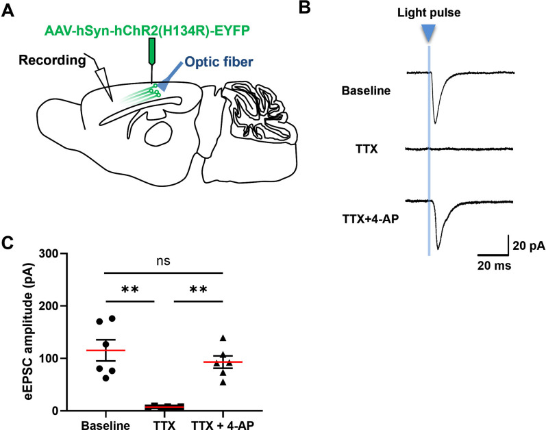
In ACC pyramidal neurons, stronger RSC stimulation evokes larger excitatory currents. Blocking AMPA receptors markedly reduces these currents; NMDA blockade or subsequent testing of the remaining kainate-receptor component does not produce a comparable additional change.
AMPA receptors receive glutamate signals and mediate fast excitatory synaptic transmission. Distinguishing them from other glutamate receptors goes beyond establishing connectivity to show how this connection primarily converts upstream activity into downstream electrical responses.
Selective activation of RSC terminals also supports direct synaptic responses, establishing a monosynaptic excitatory connection. Different ACC inputs need not share identical receptor compositions; this route illustrates a predominantly AMPA-mediated form of transmission.
Source: Hao S et al. · PLoS biology · CC BY
Original figure and English caption ↗
Weaker mechanical stimulation elicits responses more readily
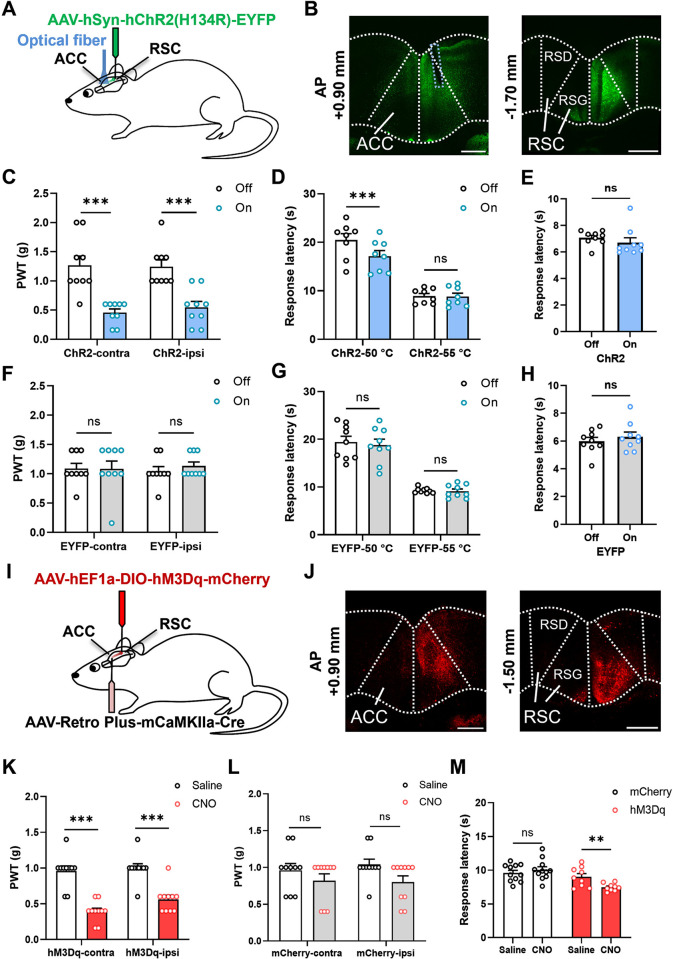
Activating RSC terminals in ACC lowers mechanical withdrawal thresholds in both hindpaws. Although the intervention is unilateral, behavioral changes occur bilaterally. Activating the cell bodies of projection-defined excitatory neurons in RSC likewise reduces both thresholds.
The response threshold shifts downward, so weaker mechanical stimulation can elicit withdrawal. Both activation approaches support a facilitatory role for this cortical connection, while the precise route producing bilateral responses may involve broader brain connectivity.
Source: Hao S et al. · PLoS biology · CC BY
Original figure and English caption ↗
Hot-plate responses accelerate while tail-flick remains similar
Thermal stimulation reveals finer distinctions. Activating RSC terminals in ACC shortens paw-lifting latency on the 50 °C hot plate without a clear change at 55 °C. Activating the cell bodies of ACC-projecting excitatory RSC neurons shortens latency at 55 °C. Effects therefore depend on activation site and stimulus condition.
In a separate tail-heating test during terminal activation, tail-flick latency remains similar. Hot-plate behavior requires supraspinal integration, whereas tail-flick emphasizes a reflex response. This distinction directs attention toward processing in the brain, particularly communication between cortical regions.
More sensitive mice do not show more avoidance
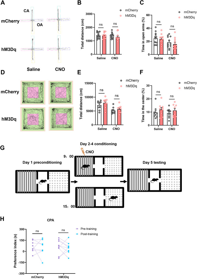
The study also examines choices between exposed and enclosed spaces. After activating these projection-defined cells, elevated-plus-maze open-arm time, open-field center time and total travel distance show no clear changes. Repeated place conditioning likewise produces no clear avoidance of the activation-paired side.
Enhanced mechanical and selected thermal responses do not come with changes in the tested emotional behaviors. Even within ACC-related networks, different inputs may emphasize different functions; the clearest change here is behavioral sensitivity to nociceptive stimulation.
Source: Hao S et al. · PLoS biology · CC BY
Original figure and English caption ↗
Another role for a memory-related region
This adult male mouse study gives the memory- and navigation-related RSC another concrete functional connection: its excitatory input can facilitate ACC-related nociceptive responses. Whether contextual memory uses this route to alter nociception is a subsequent question; the experiments directly establish connectivity, synaptic transmission and behavior during acute stimulation. It shifts attention from the physical stimulus alone to how communication between cortical areas adjusts responsiveness, offering a basis for comparing how different inputs emphasize nociception and emotion.