Why do pain and mood changes overlap after thalamic hemorrhage? A story revealed by lesion maps
Starting with lesion observations in twelve patients, a study follows posterior thalamus in mice and finds location-, time- and side-related differences in sensitivity, later behavior and neural responses.
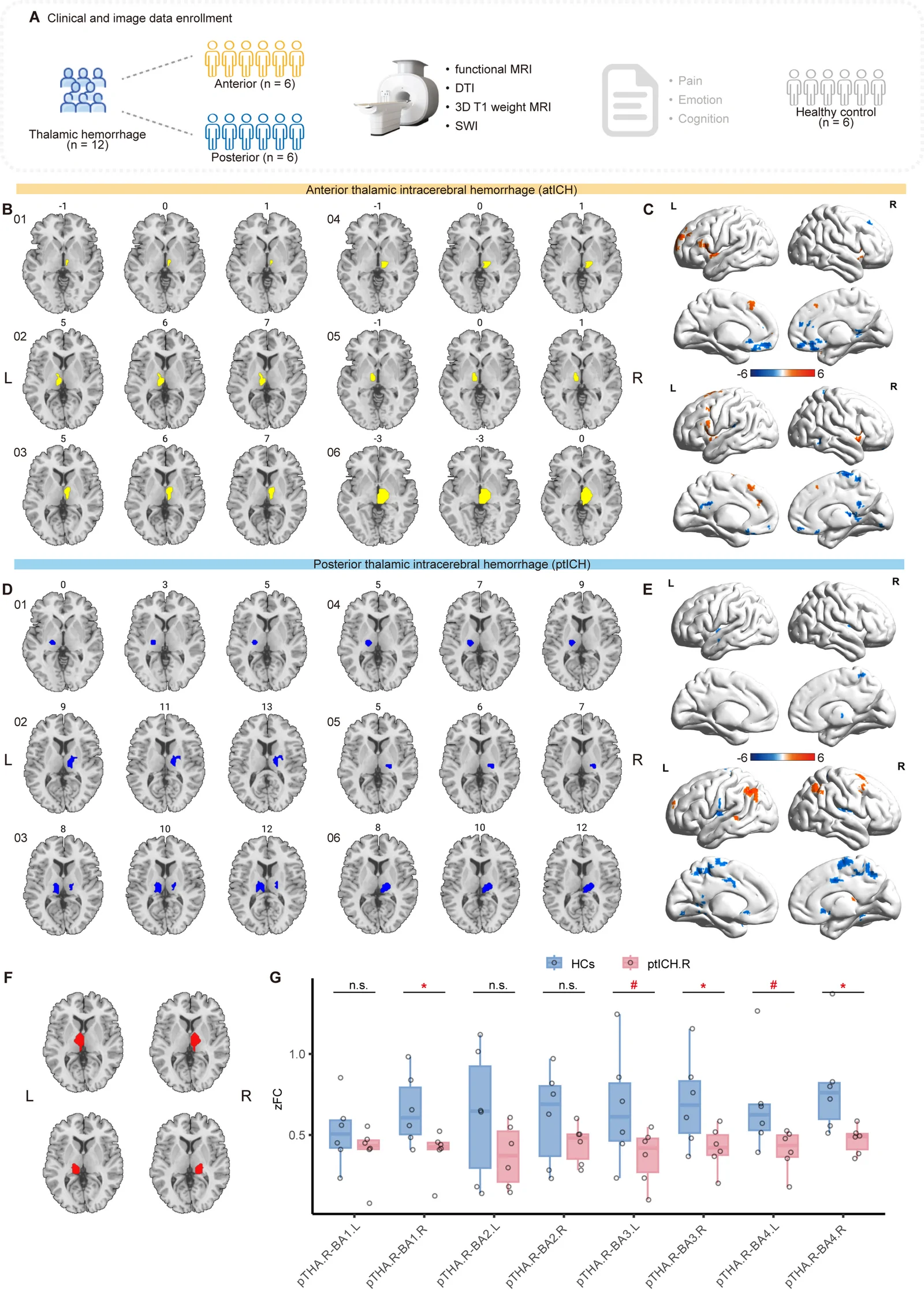
Recovery after thalamic hemorrhage can involve pain and mood symptoms as well as movement. Why do these difficulties occur together, and what does lesion location change? A 2026 BMC Medicine study maps patient lesions and then examines individual mouse thalamic nuclei. One posterior nucleus relates to persistent mechanical sensitivity and later behavioral changes, with different patterns emerging on the left and right.
After posterior-nucleus hemorrhage in mice, mechanical sensitivity appears early and selected affective behaviors change later, with side-related differences. Clinical imaging and cortical projections provide connected network clues.
Contents · 7 sections
Anterior and posterior lesions accompany different symptom patterns
The study included twelve patients with isolated thalamic hemorrhage, six anterior and six posterior. Pain, anxiety and depression scores were more prominent in the posterior group. Resting-state imaging also showed altered thalamic–sensorimotor connectivity, particularly when examining the network from the right posterior thalamic region.
These patients were at different recovery stages: median assessment intervals were about forty months for anterior cases and fifteen for posterior cases. Symptom–connectivity correlations were nonsignificant. The clinical observations offered a location clue, prompting comparisons of focal injuries in mice to resolve the relevant nucleus.
Source: Jin Y et al. · BMC medicine · CC BY-NC-ND
Original figure and English caption ↗
From the broader thalamus to posterior nucleus PO
The thalamus contains nuclei with different connections rather than one uniform relay. Mouse experiments compared extensive hemorrhage with focal injury to PO, PF and VPL. Extensive and focal PO injury made the contralateral hindpaw withdraw more readily from mechanical stimulation; neighboring and anterior controls lacked the same change.
Mechanical sensitivity persisted from early stages to around day twenty-three. Focal PO injury reproduced this aspect of extensive injury, making location closely relevant to the outcome. Mouse localization refined the clinical posterior-thalamic clue: imaging identified a broader region, while focal models examined its internal organization.
Mechanical sensitivity appears before selected behavioral changes
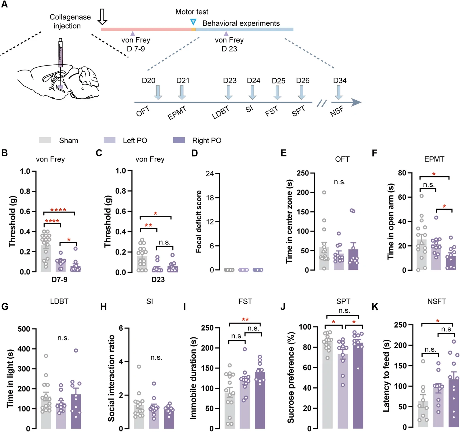
After right extensive or focal PO hemorrhage, mechanical thresholds fell early, while forced-swim immobility increased only later. Motor testing before behavioral assessment showed broadly recovered mobility comparable to controls. Sensory responses and later coping behavior therefore had different onset times.
Later changes did not span every task. Several open-field, light–dark, social and sucrose measures remained similar, while open-arm time and latency to eat in a novel setting changed. Each assay captured one facet, and following them through time distinguished changes that traveled together from preserved behavior.
The same nucleus gives different patterns after left and right injury
Both left and right PO hemorrhage lowered contralateral mechanical thresholds, with a stronger acute effect on the right. Chronically, left injury mainly reduced sucrose preference, while right injury increased swim immobility and selected avoidance measures. The nearly symmetric thalamic sides produced different behavioral patterns.
The patterns also retained overlap: some direct left–right comparisons were nonsignificant, and several social, open-field and light–dark behaviors remained unchanged. Laterality gave selected measures a bias rather than dividing sensation and affect into two separate boxes, guiding the next neural observations.
Source: Jin Y et al. · BMC medicine · CC BY-NC-ND
Original figure and English caption ↗
After injury, a weak stimulus evokes stronger activity
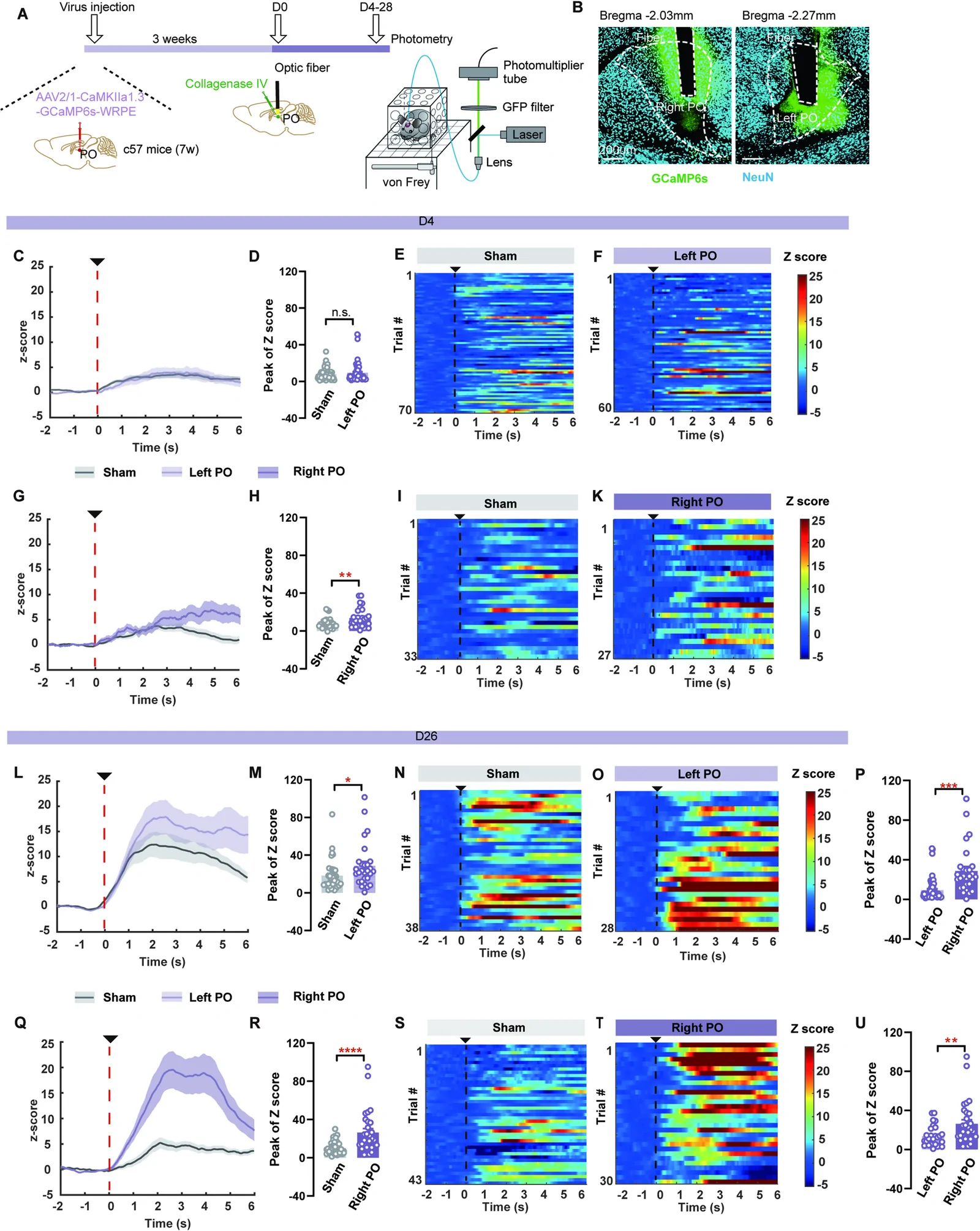
Tissue damage need not simply quiet the surviving network. PO population calcium responses to weak mechanical stimulation increased acutely after right injury, without the same significant left effect. Chronically, both sides showed stronger responses than their earlier stages, revealing changing network reactions to the same stimulus.
The enhancement depended on context: acute footshock increased right-side activity, while tail suspension and restraint did not reproduce it. Neural responses paralleled sensitivity and later behavior over time, although recording alone did not identify the cell or output driving behavior. It added network dynamics to the static lesion map.
Source: Jin Y et al. · BMC medicine · CC BY-NC-ND
Original figure and English caption ↗
Different output patterns add structural context
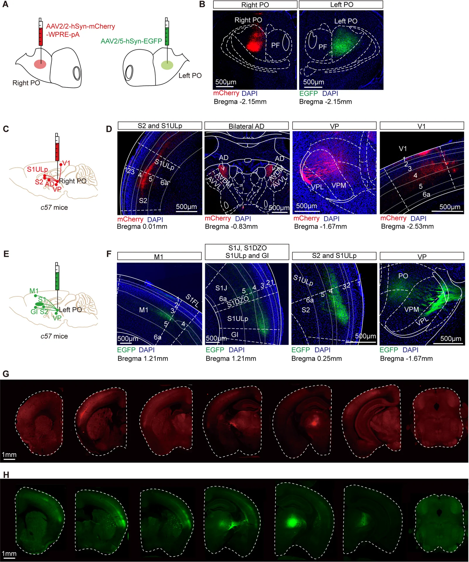
Tracing suggested different left and right PO labeling patterns in sensory, motor and other regions, with cortical outputs mainly ipsilateral. These connections provide structural context for side-related behavior and place a local injury within a broader network account.
The connections remain candidate explanations, with specific output contributions awaiting selective functional tests. The findings already bring nucleus location, side, evolving neural responses and task-specific behavior onto one map, giving symptom co-occurrence more concrete spatial and temporal coordinates.
Source: Jin Y et al. · BMC medicine · CC BY-NC-ND
Original figure and English caption ↗
Recovery has both a location and a timeline
From twelve patients to focal hemorrhage in male mice, the study turns a broad pairing of post-thalamic-hemorrhage pain and mood changes into a more detailed process. The clinical observations require larger, time-matched cohorts, while different mouse tasks separately inform sensation, reward preference and coping behavior.
Lesion location does not tell the entire story: side and recovery stage change the responses and behaviors observed. Putting these coordinates together helps explain how an injured network continues functioning and influences different aspects of recovery.