Why can light touch evoke a stronger response? A sensory-amplifying loop between cortex and thalamus
A mouse nerve-injury study identifies a connection that leaves sensory cortex, passes through thalamus and returns to cortex, amplifying responses to weak mechanical stimulation with different effects in normal and injured states.
Why can the same light touch evoke a stronger response after nerve injury? The stimulus may be unchanged while its processing in the brain has shifted. Sensory information travels through thalamus to cortex, and cortex sends signals back. This PAIN study follows that return route in mice: from the hindlimb region of primary somatosensory cortex (S1HL) through ventral posterolateral thalamus (VPL), then back to the same cortical region. The connection particularly enhances responses to weak mechanical stimulation, linking light touch and hypersensitivity through a specific feedback network.
S1HL–VPL–S1HL forms a predominantly excitatory return loop. Increasing its activity amplifies weak-stimulus responses; reducing activity attenuates mechanical hypersensitivity and spontaneous pain-like behavior after nerve injury while largely preserving normal baseline thresholds.
Contents · 6 sections
Sensory cortex becomes more active alongside hypersensitivity
In the nerve-injury model, activity-related Fos labeling in S1HL increased at both seven and thirty days, mostly in neurons carrying an excitatory-cell marker. These cells project to VPL and posterior thalamus, with fewer descending projections from the corresponding inhibitory population. The cortical change had an output the researchers could follow.
A more active region might receive stronger input or help amplify it. The team turned to VPL and compared cortical signals with ascending spinal input. Did they reach the same cells? The question moved the work from regional activity toward the flow of information through the network.
One thalamic nucleus receives distinct sources of information
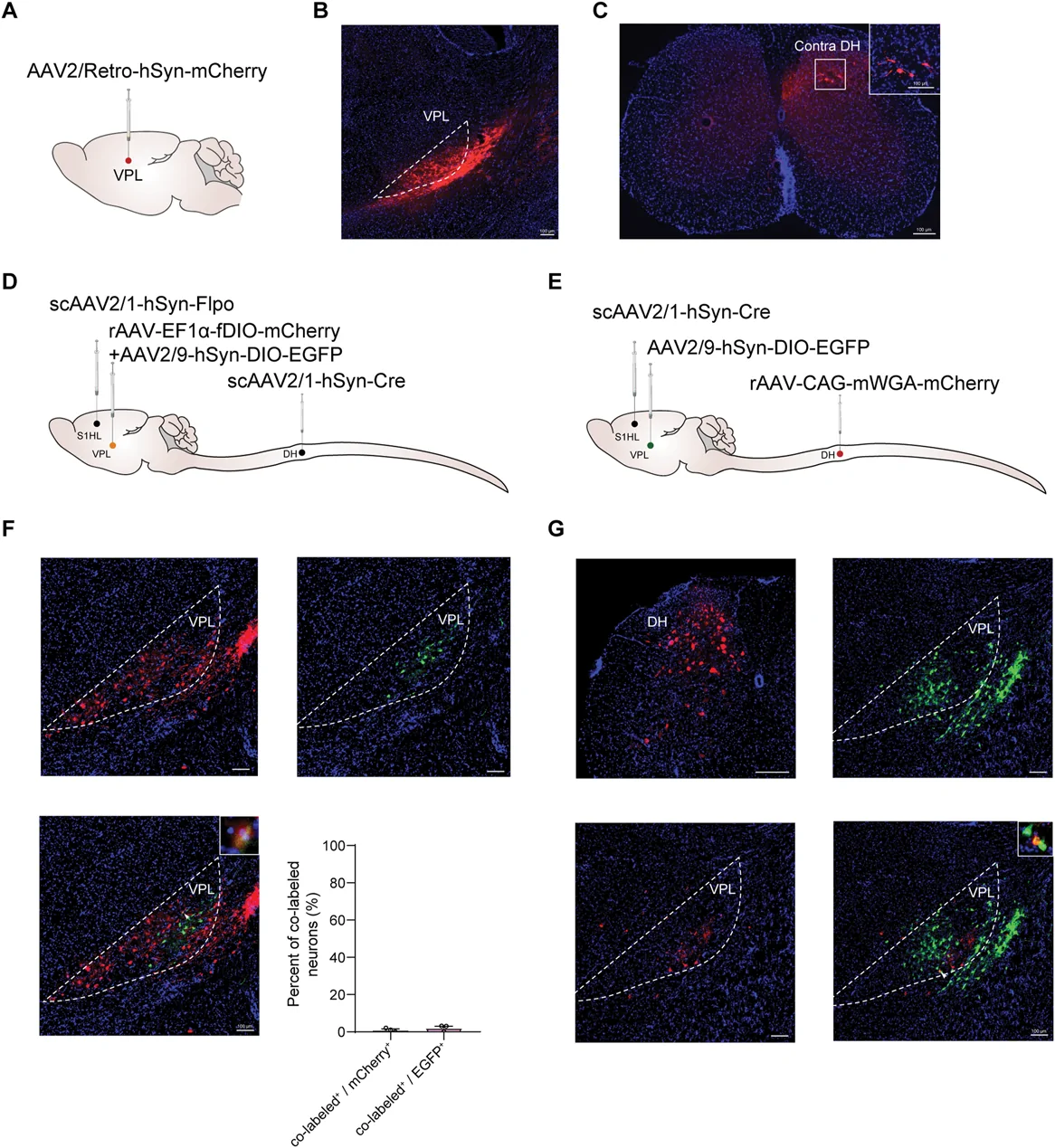
VPL is often viewed as a somatosensory relay, and the study traced input from contralateral lumbar dorsal horn. Yet VPL cells receiving S1HL input showed little spatial overlap with those receiving spinal input. Incoming information entered through different routes rather than converging entirely on one population.
During mechanical stimulation in nerve-injured mice, the VPL population receiving cortical input showed stronger calcium responses. A difference in input source was thus linked with altered activity after injury. These populations are defined by their connections; their detailed cellular identities remain a separate question.
Source: Hu Y et al. · Pain · CC BY-NC-ND
Original figure and English caption ↗
The same connection behaves differently across states
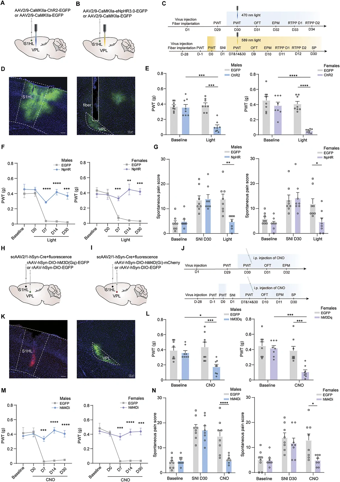
Increasing S1HL terminal activity in VPL, or activity of the receiving VPL population, lowered mechanical withdrawal thresholds in both male and female mice. Reducing activity instead attenuated post-injury mechanical hypersensitivity and spontaneous pain-like behavior in later observations.
Normal baseline thresholds did not change clearly with inhibition, showing that the connection’s effects depend on state. Open-field, elevated-plus-maze and place-preference observations did not show corresponding affect-related improvements. Attenuated mechanical responses occurred without every measured behavior changing alongside them.
Source: Hu Y et al. · Pain · CC BY-NC-ND
Original figure and English caption ↗
A cortical signal finds its way back to cortex
VPL cells receiving S1HL input sent much of their output back to sensory cortex, with denser terminals in layer IV. Most recipient cells carried excitatory markers, alongside a small inhibitory fraction. A route beginning in cortex therefore re-entered its local network.
This return structure allows cortex to receive information and influence its own responses through thalamus. Activity on both sides also synchronized during unilateral hindpaw stimulation, with interhemispheric connections potentially contributing to propagation. The local loop operates within a wider bilateral network.
The amplification favors weaker inputs
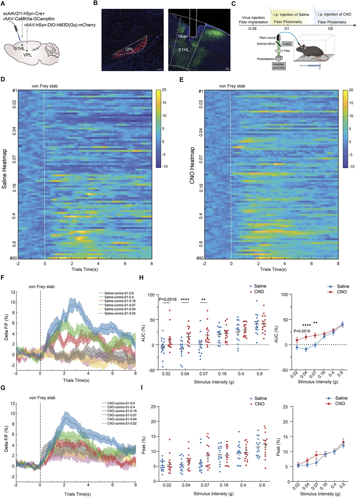
A revealing difference appeared across stimulus strengths. Increasing cortical-input-defined VPL activity in normal mice enhanced S1HL calcium responses to low-intensity mechanical stimulation, while corresponding differences at higher intensities were nonsignificant. Female mice showed a similar pattern. The feedback did not affect every stimulus equally.
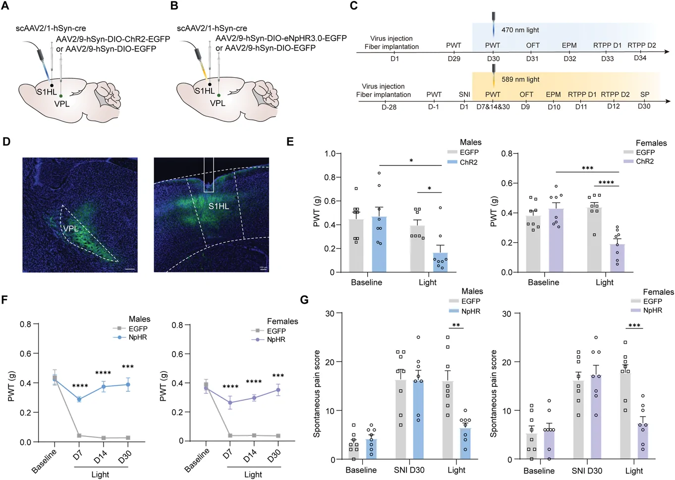
Reducing activity after injury attenuated the abnormal cortical response to weak stimulation. Changing VPL terminals returning to S1HL also influenced mechanical hypersensitivity and spontaneous pain-like behavior. The anatomical return route and functional outcomes converge on its contribution to pathological amplification of weak input.
Source: Hu Y et al. · Pain · CC BY-NC-ND
Original figure and English caption ↗
Sensory responses are shaped by return traffic
Response strength therefore depends on more than the signal entering the brain. Return connections between cortex and thalamus can also shape the response to weak input. This study links post-injury mechanical hypersensitivity with a feedback loop distinguished by input source, state and stimulus intensity.
The evidence concerns mouse nerve injury, mechanical stimulation and associated pain-like behavior. The new perspective is that sensory cortex is more than an endpoint: it helps shape information that subsequently returns to it. Understanding hypersensitivity involves both ascending transmission and this return processing.
Source: Hu Y et al. · Pain · CC BY-NC-ND
Original figure and English caption ↗