An endogenous brainstem brake: an enkephalin-related pathway regulates sensitivity
From baseline thresholds to inflammation and nerve injury models, an RVM→SRD connection offers cellular clues to brainstem regulation of pain.
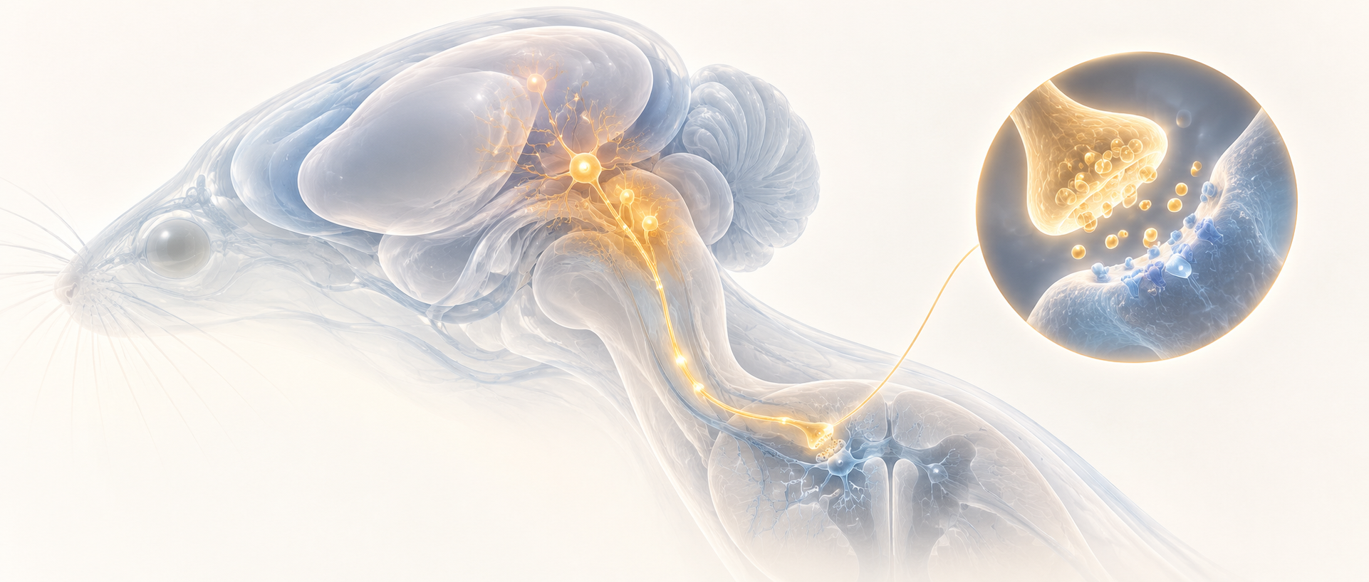
As pain-related information travels toward the brain, the brainstem is more than a passive relay. Its networks also regulate sensory input: some populations facilitate hypersensitivity, while others inhibit it. How can a defined regulator be identified within these mixed populations? A 2026 CNS Neuroscience & Therapeutics study examines enkephalin-related neurons in the rostral ventromedial medulla, or RVM, and follows their projection to the subnucleus reticularis dorsalis, or SRD. Opposing effects of activation and suppression reveal a connection involved in baseline sensitivity and modulation in two pain models.
Enkephalin-related RVM neurons and their SRD projection contribute to baseline antinociceptive regulation. Increasing their activity raises baseline thresholds and attenuates hypersensitivity in inflammatory and nerve-injury models.
Contents · 6 sections
These cells are recruited during hypersensitivity
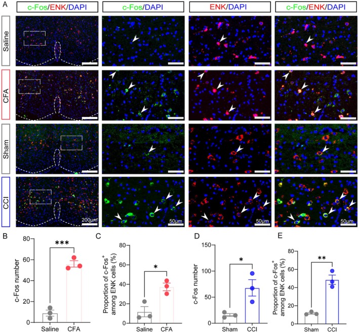
In CFA inflammation and CCI nerve-injury models, mice had lower mechanical and thermal withdrawal thresholds. RVM c-Fos-positive cells increased, including more activity labeling within the enkephalin-related population. Recruitment during persistent pain prompted investigation of how these cells participate in sensory regulation.
RVM contains functions with different directions of influence, and increased activity during pain could reflect feedback countering persistent input. Penk-based genetic labeling identified the enkephalin-related cells for direct activity modulation, linking molecular identity with behavioral function. The activity markers thus led to functional tests.
Source: Wan K et al. · CNS neuroscience & therapeutics · CC BY
Original figure and English caption ↗
Activation and suppression shift thresholds in opposite directions
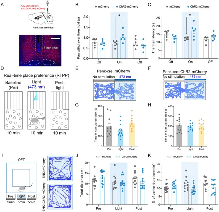
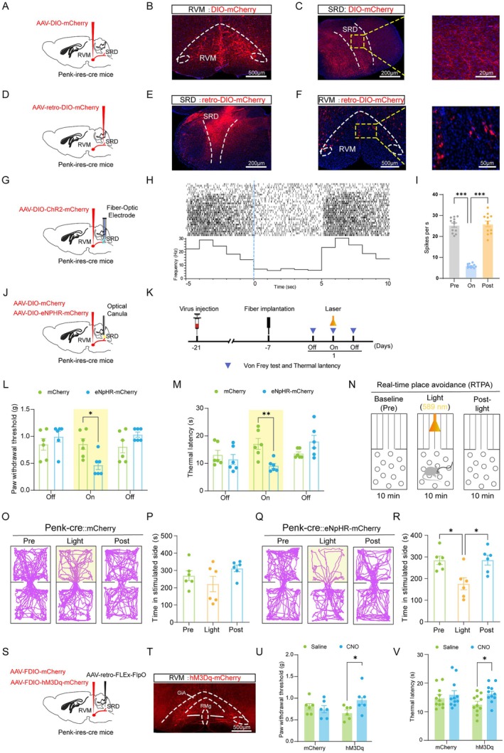
In naïve mice, chemogenetic suppression lowered mechanical and thermal withdrawal thresholds. Optical suppression likewise produced rapid, reversible hypersensitivity. Increasing activity raised baseline thresholds. Convergence across strategies showed that these cells contribute to sensory regulation even without injury.
Locomotor distance and open-field anxiety-related measures remained similar. Mice avoided the paired location during brief suppression, whereas groups did not differ significantly in the longer-term conditioned-place assay. Conversely, activation raised thresholds in naïve mice without making them prefer the paired location. The population’s influence on sensory stimulation and place choice thus varied with timing and state.
Source: Wan K et al. · CNS neuroscience & therapeutics · CC BY
Original figure and English caption ↗
The same activation produces place preference during pain
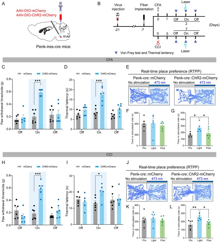
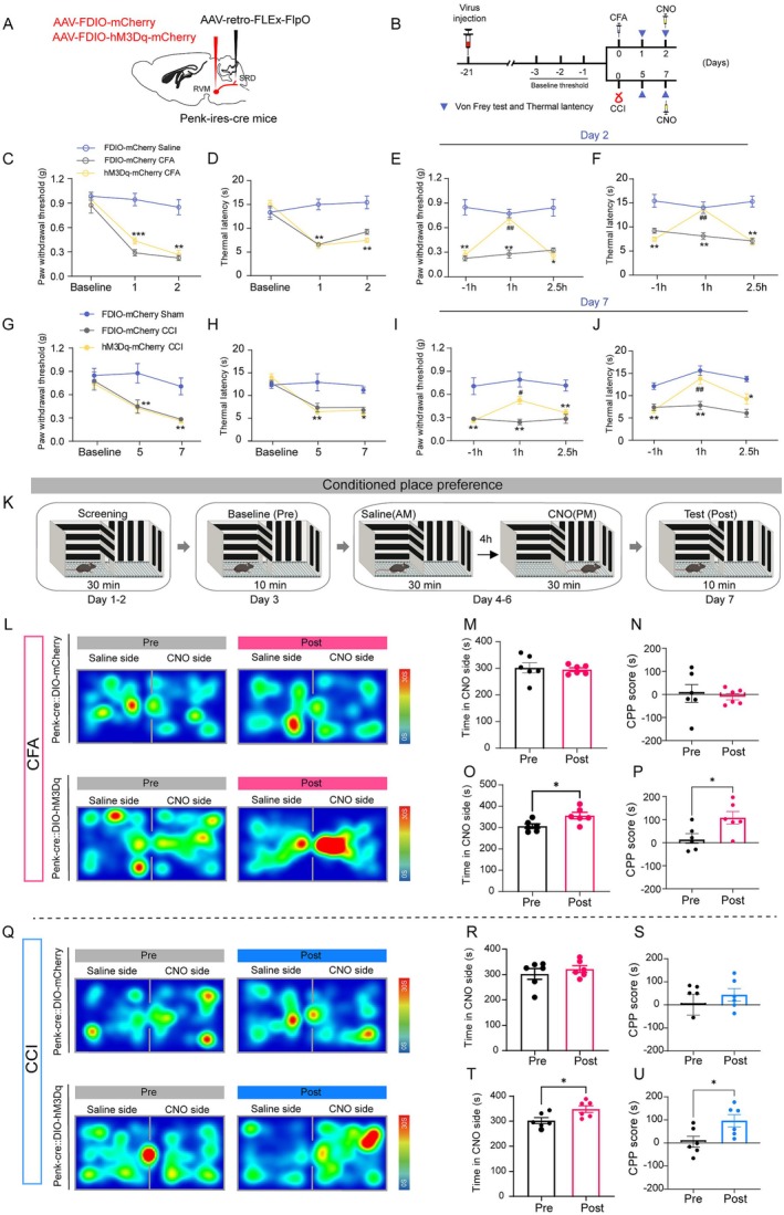
In CFA and CCI models, increasing RVM population activity raised mechanical and thermal thresholds, and animals spent more time in the stimulation-paired location. This preference was consistent with relief-related motivation during ongoing pain. Differences between naïve animals and pain models revealed a state-dependent relationship between brainstem modulation and behavioral choice.
The same cellular modulation can have different motivational outcomes in different states. Chemogenetic effects returned toward baseline after roughly two and a half hours, showing a reversible time course. Thresholds and place choices together describe sensory and motivational responses that can be changed over the short term during pain.
Source: Wan K et al. · CNS neuroscience & therapeutics · CC BY
Original figure and English caption ↗
A subset of RVM cells sends its output to SRD
Bidirectional tracing confirmed the RVM–SRD connection. SRD-projecting cells made up about 35.06% of the enkephalin-related RVM population. Increasing terminal activity in SRD suppressed target-neuron firing. Molecular identity, anatomy and local firing changes focused the investigation on this output as a defined site of inhibitory brainstem regulation.
Suppressing the relevant SRD terminals lowered baseline mechanical and thermal thresholds and accompanied real-time avoidance. Increasing SRD-projecting cell activity raised baseline thresholds, with preserved locomotor and open-field measures. Convergent findings from the wider population and this output identify RVM→SRD as an important connection for understanding baseline antinociceptive regulation.
Source: Wan K et al. · CNS neuroscience & therapeutics · CC BY
Original figure and English caption ↗
Enhancing SRD-projecting cells attenuates hypersensitivity
Increasing SRD-projecting cell activity raised mechanical and thermal withdrawal thresholds in both CFA and CCI models and produced conditioned preferences consistent with relief. Convergence across the two models showed regulatory capacity in different injury contexts.
Effects returned after roughly two and a half hours, paralleling reversible modulation of the broader RVM population. Existing suppression experiments mainly described baseline function, while activation tests extended the connection to pain models. Suppression during pain will help clarify how much compensation is provided by the recruitment already present after injury.
Source: Wan K et al. · CNS neuroscience & therapeutics · CC BY
Original figure and English caption ↗
Adding cellular identity and connectivity to brainstem pain regulation
The study adds a defined cellular population and output target to brainstem antinociceptive regulation. Enkephalin-related identity also provides a molecular entry point for investigating peptide release, cotransmitters and receptors within SRD. Convergent clues from target firing and behavior provide a foundation for connecting these levels.
The results come from adult male mice. Studies across sexes, other output collaterals and longer timescales will extend the picture. The findings show that cellular recruitment during pain can include regulatory capacity that remains available for enhancement, turning an endogenous brake into a more concrete circuit question.