Visual cortex also shapes pain sensitivity: two thalamic projections, different effects
A male-mouse study finds greater V2M excitatory-cell activity after nerve injury; inhibiting terminals projecting to thalamic LP eases hypersensitivity, while LD-projecting terminals lack the same effect.
Visual cortex brings sight to mind, but some of its cells also participate in processing bodily nociceptive information. A study published on March 11, 2025 in Communications Biology examines medial secondary visual cortex, V2M. Its glutamatergic cells become more active after nerve injury in male mice, and suppressing them reduces mechanical and heat hypersensitivity. Following the outputs to the thalamus reveals different effects in two innervated nuclei: LP contributes to pain-sensitivity regulation, whereas LD lacks the same effect under the tested conditions.
V2M glutamatergic cells and their LP projection contribute to pain-sensitivity regulation, with effects depending on injury state and downstream target.
Contents · 7 sections
V2M activity increases after nerve injury
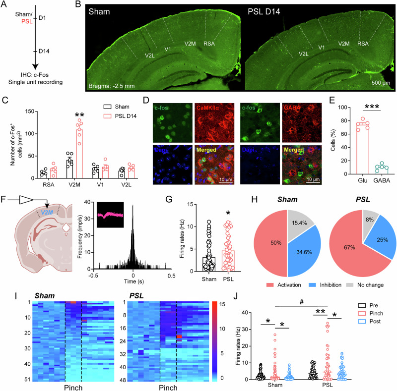
After partial sciatic-nerve injury, activity-marker-positive cells increase in V2M, without the same clear change in tested neighboring regions such as primary visual cortex and lateral secondary visual cortex. Most increased markers overlap glutamatergic cells, focusing the study on this excitatory output.
Single-unit recordings show more frequent spontaneous firing and stronger responses to noxious hindpaw stimulation, with a larger fraction of recorded cells activated. Some cells are suppressed or lack a clear response, revealing heterogeneity within the region.
Source: Tan B et al. · Communications biology · CC BY-NC-ND
Original figure and English caption ↗
Several hindpaw stimuli evoke stronger population signals
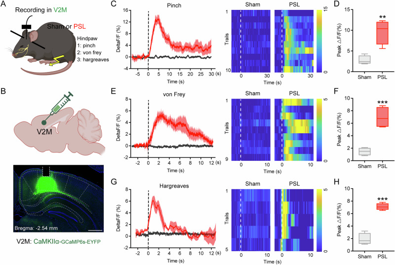
Population calcium recordings from V2M glutamatergic cells in awake mice show stronger responses to pinch, mechanical and heat stimuli after nerve injury. Control mice lack the same clear population changes, relating nociceptive input to cortical responses in living animals.
Population calcium and single-unit recordings have different scopes: one aggregates cellular activity while the other resolves individual firing. Both show stronger post-injury responses, and single-cell diversity helps explain why control population changes need not be clear.
Source: Tan B et al. · Communications biology · CC BY-NC-ND
Original figure and English caption ↗
Changing one cortical side affects both hindpaws
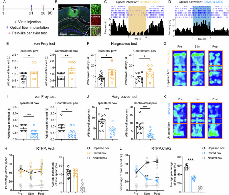
In uninjured mice, suppressing cells in one V2M raises mechanical thresholds and heat-withdrawal latencies in both hindpaws. Activating the cells lowers those measures and induces avoidance of the activation-associated location. Higher thresholds mean that stronger mechanical stimulation is needed for withdrawal, while longer heat latencies indicate later withdrawal. Together, the two stimulus types show bilateral sensitivity effects of a unilateral cortical change.
Although suppression changes sensitivity measures, it does not induce a clear place preference. Sensory-response changes and place choice do not always move together: baseline suppression differs from activation-associated avoidance.
Source: Tan B et al. · Communications biology · CC BY-NC-ND
Original figure and English caption ↗
Suppression helps after injury, further activation adds no clear worsening
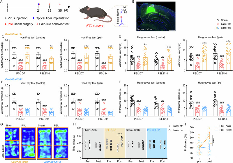
After nerve injury, V2M suppression improves mechanical and heat hypersensitivity. Mice also prefer the suppression-associated location, unlike the absent baseline place preference, suggesting relief of an injury-associated aversive component.
Further activation of the already more active V2M does not clearly lower thresholds further or alter place choice. Different outcomes across states show context-dependent regulation rather than unlimited amplification of sensitivity.
Source: Tan B et al. · Communications biology · CC BY-NC-ND
Original figure and English caption ↗
Two thalamic nuclei receive projections
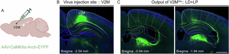
Tracing reveals dense glutamatergic V2M projections to the lateral posterior nucleus, LP, and laterodorsal nucleus, LD. Cortical–thalamic communication is not limited to ascending sensory information; cortical outputs can influence downstream processing.
Axon presence alone does not identify its behavioral contribution. The study therefore changes V2M terminal activity in each nucleus and measures sensitivity, bringing the two anatomical connections into a functional comparison.
Source: Tan B et al. · Communications biology · CC BY-NC-ND
Original figure and English caption ↗
LP terminal manipulation has effects absent at LD
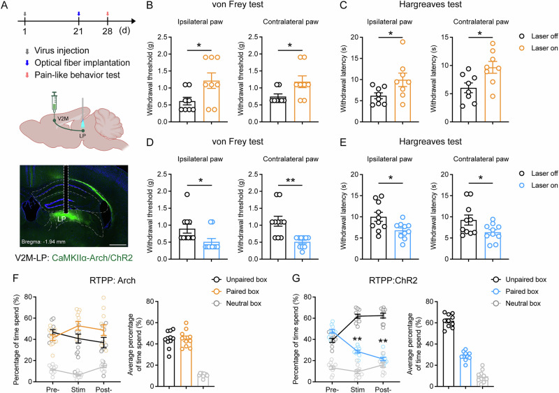
In uninjured mice, inhibiting V2M terminals in LP raises mechanical thresholds and heat latencies; activation lowers both measures and induces place avoidance, reproducing part of the cell-body effects. LP-terminal inhibition still lacks a clear place preference.
Comparable activation or inhibition of LD terminals does not clearly change tested sensitivity in either hindpaw. Neighboring nuclei that both receive projections have different effects in these assays, highlighting downstream target identity.
Source: Tan B et al. · Communications biology · CC BY-NC-ND
Original figure and English caption ↗
LP inhibition still provides relief, with different LD results
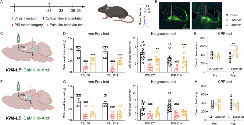
In nerve-injured mice, suppressing LP terminals improves bilateral mechanical and heat sensitivity and produces suppression-associated place preference. LD-terminal inhibition does not clearly change sensitivity or place choice, extending the projection difference into the injury state.
This male-mouse study places a specific visual-cortical population within a pain-sensitivity network and shows how state and target shape its effects. How V2M integrates visual and bodily information, and which LP cells carry the influence onward, remain specific next questions.
Source: Tan B et al. · Communications biology · CC BY-NC-ND
Original figure and English caption ↗