Up from spinal cord and back from brain: how is mechanical hypersensitivity maintained?
Parallel thalamic inputs engage somatosensory cortex, lateral superior colliculus and descending medullary cells; repeated activation produces lasting hypersensitivity in uninjured mice.
After injury, relatively weak mechanical stimuli can evoke persistent hypersensitivity. Sensory signals travel toward the brain, while descending signals return to spinal cord. Can these opposing flows jointly maintain post-injury changes? A Nature study traces and manipulates spinal, thalamic, cortical, collicular and medullary routes, identifying interdependent long-range pathways. Repeated activation also produces lasting mechanical hypersensitivity in uninjured mice, whereas a single activation does not.
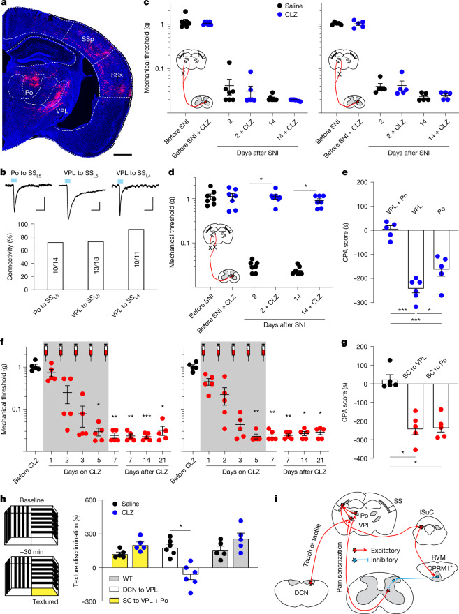
Selected ascending and descending nodes contribute to post-injury mechanical hypersensitivity. VPL and Po thalamic branches can compensate for one another; joint inhibition blocks model hypersensitivity and stimulus aversion.
Contents · 8 sections
Finding medullary cells that return to spinal cord
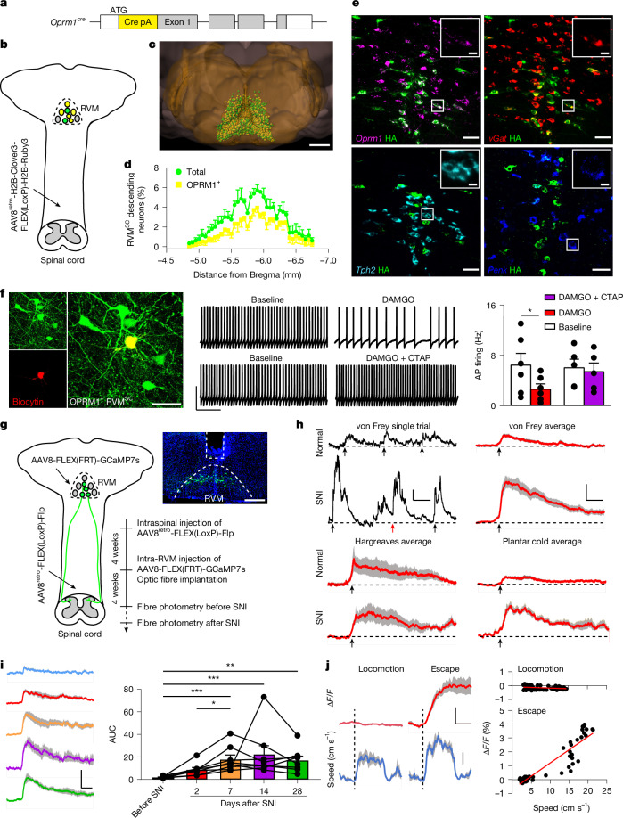
Combining Oprm1 labeling with spinal retrograde tracing identifies μ-opioid-receptor-positive RVM cells projecting to spinal cord. Most are GABAergic, with little serotonergic or Penk expression, and their axons branch toward superficial dorsal horn. Projection identity narrows a heterogeneous region to a defined descending population.
The cells show calcium responses during withdrawal, escape and spontaneous paw flinching, unlike ordinary locomotion. SNI increases mechanical and cold responses without equivalent heat increases, while CFA responses decline over time. Activity differs with sensory modality and model progression.
Source: Wang Q et al. · Nature · CC BY-NC-ND
Original figure and English caption ↗
One activation and a week of activation differ
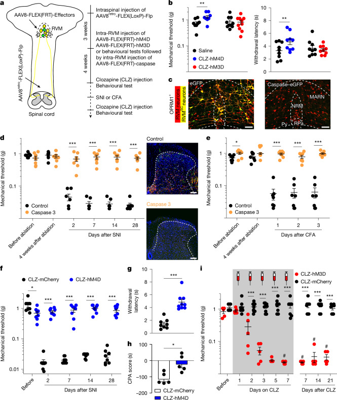
In healthy mice, acute activation does not significantly change mechanical or cold measures; inhibition produces small but significant increases in threshold and cold latency. Seven days of activation instead induces persistent mechanical and cold hypersensitivity without significant heat changes. The activity history matters.
Repeated activation strengthens mechanically evoked avoidance without increasing spontaneous licking, flinching or grimace scores. Evoked hypersensitivity and spontaneous pain-related behavior do not necessarily emerge together.
Source: Wang Q et al. · Nature · CC BY-NC-ND
Original figure and English caption ↗
The population contributes to establishment and maintenance
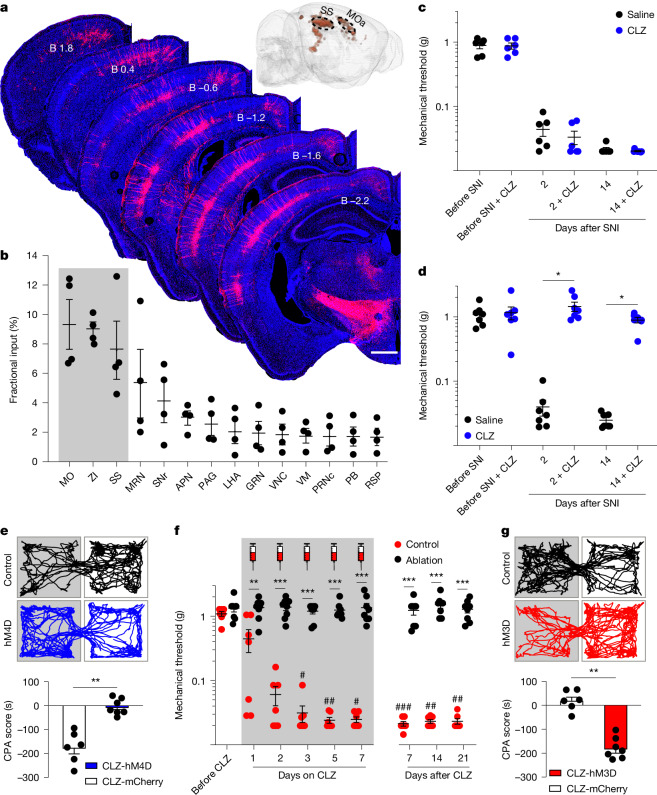
Pre-injury ablation prevents normal development of SNI and CFA mechanical hypersensitivity. Post-injury inhibition improves SNI mechanical and cold responses and reduces stimulus aversion, spontaneous behavior and grimace. Mechanical improvement remains possible at day twenty-eight, indicating contributions to both establishment and maintenance.
Affect-related measures differ. Repeated activation in healthy mice produces anxiety-like exploration changes without corresponding immobility effects. Inhibition six weeks after SNI does not significantly improve established anxiety-like or immobility measures. Sensory improvement does not automatically reverse these later changes.
Lateral superior colliculus sends excitatory input
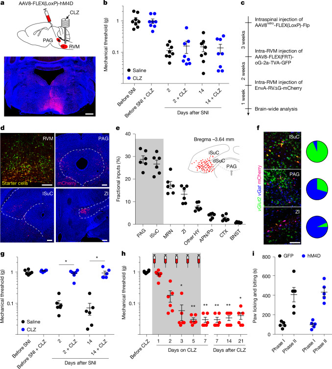
Inhibiting the tested excitatory periaqueductal-gray terminals does not significantly change healthy mechanical thresholds or SNI hypersensitivity. Lateral superior colliculus, or lSuC, supplies predominantly excitatory input. Inhibiting lSuC–RVM restores model mechanical thresholds; repeated activation induces persistent hypersensitivity in healthy mice.
The route is selective: inhibition does not similarly remove CFA heat hypersensitivity or significantly reduce formalin licking. Activation does not clearly alter tested locomotion, heart rate or breathing measures. Mechanical changes occur without broad changes across these outcomes.
Source: Wang Q et al. · Nature · CC BY-NC-ND
Original figure and English caption ↗
Cortical effects require the descending output
RVM-projecting lSuC cells receive somatosensory and premotor inputs. Somatosensory-route inhibition reduces SNI mechanical hypersensitivity and place aversion, unlike the premotor route. Repeated somatosensory activation induces hypersensitivity only when OPRM1-positive RVM spinal-projecting cells remain intact.
Source: Wang Q et al. · Nature · CC BY-NC-ND
Original figure and English caption ↗
One thalamic branch can compensate for the other
Spinally associated input reaches somatosensory cortex through VPL and Po, both influencing collicular-projecting cortical cells. Inhibiting either branch alone leaves hypersensitivity and aversion intact; joint inhibition attenuates both. Repeated activation of either branch also induces RVM-dependent hypersensitivity, demonstrating parallel functional redundancy.
Source: Wang Q et al. · Nature · CC BY-NC-ND
Original figure and English caption ↗
Texture discrimination and hypersensitivity can dissociate
Joint spinal–VPL/Po inhibition preserves tested floor and object texture preferences. By contrast, inhibiting dorsal-column-nucleus input to VPL alters texture discrimination without significant acute-response or SNI-hypersensitivity changes. Different ascending sources support distinct aspects of sensory processing.
Long-range pathways reveal how hypersensitivity persists
The study in mice of both sexes connects ascending sensation with descending modulation and reveals consequences of repeated activity. GABAergic RVM cells may promote hypersensitivity through spinal disinhibition, though the local gate cells remain unidentified. Functionally supported long-range relationships show that persistent hypersensitivity depends on both ascending signals and what the brain sends back.