Synaptic cargo and persistent pain: a new clue from glial precursor cells
In mice with bone-cancer pain, researchers found more synaptic material inside glial precursor cells in the anterior cingulate cortex. Following LCN2 signaling reveals a cellular interaction behind these changes.
Why does persistent pain so often come with unease? Clues may lie both in circuits that process sensation and emotion and in the less familiar cells beside neurons. Oligodendrocyte precursor cells (OPCs) are known for generating myelinating cells, but they also engulf nearby synaptic material. A 2026 study in Acta Neuropathologica Communications follows this interaction in the anterior cingulate cortex (ACC) of mice with bone-cancer pain, connecting changes in brain activity, material inside glial cells and a molecular signal.
In the bone-cancer-pain model, ACC OPCs contained more inhibitory-neuron-associated material. Reducing LCN2 or its receptor SLC22A17 decreased this material and improved mechanical sensitivity and selected anxiety-like measures, highlighting signaling between neurons and glial cells.
Contents · 6 sections
How does the brain’s inhibitory brake respond to persistent pain?
By day 21, male model mice were more sensitive to mechanical stimulation and entered the center of an open field and the open arms of an elevated maze less often. ACC calcium responses to weak stimulation and activity-related labeling increased alongside these behavioral changes, making this region a starting point for tracing cellular alterations.
GAD67-positive cell counts and inhibitory synaptic markers fell, yet the recorded GABA neurons responded more strongly. The researchers interpret the stronger response as compensation within a structurally altered inhibitory network. Enhancing these neurons’ activity reduced mechanical sensitivity and improved selected exploration measures, linking their activity to behavioral regulation.
A closer look reveals synaptic material inside glial cells
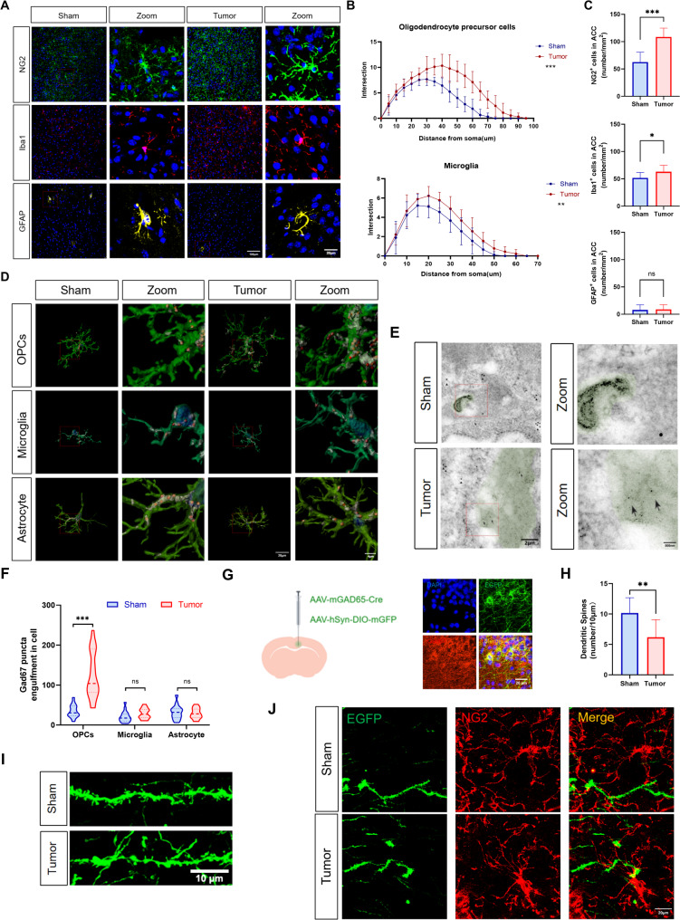
The team next turned from neurons to their neighbors. OPCs had more branches in model mice, and 3D reconstructions showed more GAD67-associated material inside them, together with lysosomal labeling. Immunoelectron microscopy provided corresponding intracellular observations. Together, the images place these precursors within the handling of inhibitory-neuron-associated material.
Dendritic spines on GABA neurons also decreased, and the team observed OPC contact with these structures. Spines receive inputs, whereas neuronal axons carry outputs. These distinct parts of the circuit help frame the findings: pain-associated remodeling involves both how neurons respond and how neighboring glial cells handle surrounding structures.
Source: Jiang L et al. · Acta neuropathologica communications · CC BY-NC-ND
Original figure and English caption ↗
Following LCN2 between cells
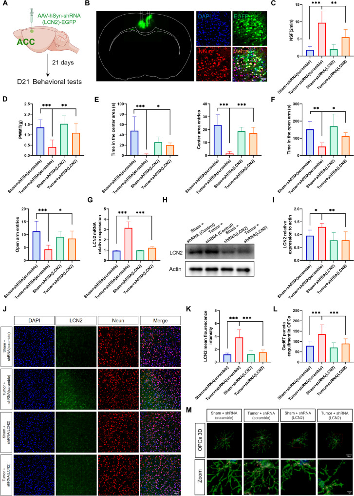
What signal connects OPC changes with the pain state? Molecular screening brought the secreted protein LCN2 into view. Its levels rose in both ACC tissue and serum, with ACC colabeling mainly associated with neurons. Brain activity, cellular morphology and molecular expression thus converged on the same candidate signal.
Neutralizing LCN2 in the ACC or reducing neuron-associated LCN2 expression improved selected pain-like and anxiety-like measures and decreased GAD67-associated material inside OPCs. Changing one molecular signal altered both intracellular material and behavior, strengthening interest in LCN2 as part of the neuron–glia interaction.
Source: Jiang L et al. · Acta neuropathologica communications · CC BY-NC-ND
Original figure and English caption ↗
The receptor side responds too
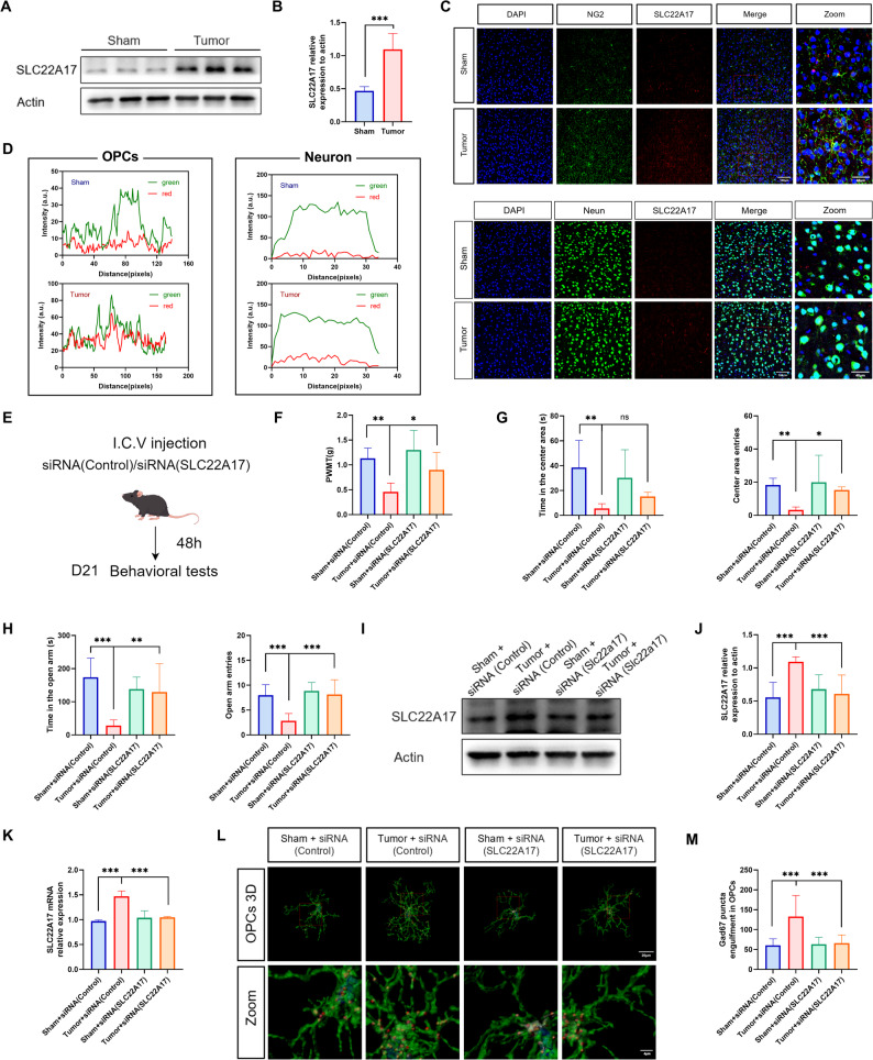
A signal also needs a receiver. SLC22A17, an LCN2 receptor, increased in the model, including stronger OPC-associated colabeling. Reducing receptor expression in the ACC improved mechanical thresholds and selected exploration measures while decreasing material inside OPCs. Interventions at the ligand and receptor ends produced consistent changes.
The LCN2–SLC22A17 axis connects molecular signaling with glial handling of cellular material. Because receptor intervention targeted local brain tissue, the contribution of individual cell classes remains an open question. The advance is a link that can be explored experimentally across molecular, cellular and behavioral changes.
Source: Jiang L et al. · Acta neuropathologica communications · CC BY-NC-ND
Original figure and English caption ↗
From receiving a signal to reshaping cellular extensions
Engulfment depends on the cell’s own machinery. LCN2 increased fluorescent-bead uptake and lysosome-related measures in cultured primary OPCs, while reducing SLC22A17 weakened these changes. Beads provided a readout of uptake capacity through which the team could explore how signaling changes cellular behavior.
The trail led to the cytoskeleton. LCN2 was associated with changes in filopodia and RAC1 activity, and blocking actin polymerization reduced bead uptake. Cellular extensions and their internal scaffolding help shape contact and uptake, linking tissue observations with a cellular mechanism explored in culture.
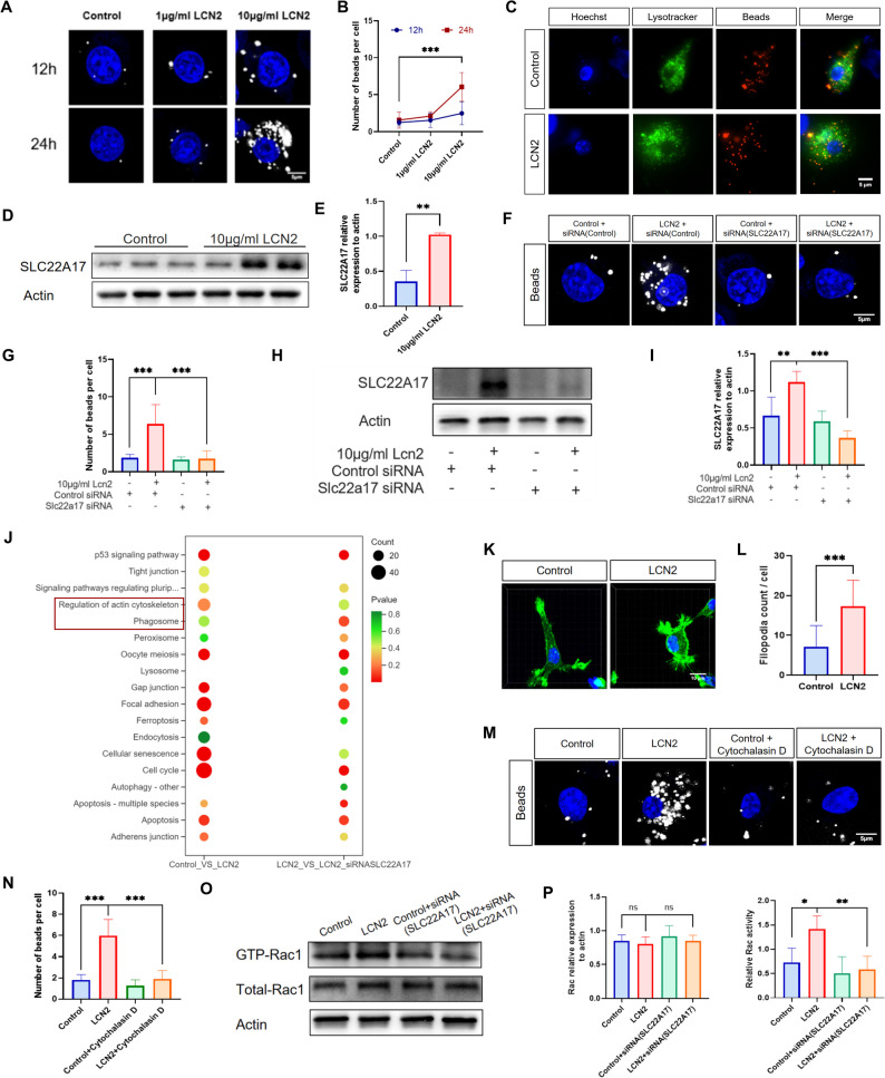
Source: Jiang L et al. · Acta neuropathologica communications · CC BY-NC-ND
Original figure and English caption ↗
Understanding pain includes looking at neurons’ neighbors
The study identifies a neuron–glia link in bone-cancer pain: ACC LCN2 signaling, material uptake by OPCs and inhibitory-network changes accompany pain-like and anxiety-like behavior. Circuit remodeling includes neuronal activity as well as structural interactions between cells, bringing glial precursors into the foreground of pain research.
The findings come from a male-mouse model. Tracking material over time, measuring synaptic currents and testing the signal in specific cell classes will help connect structural changes to circuit function. Other sexes, tumor types and clinical settings also await study. Following this synaptic material may reveal more about why pain and emotional changes become intertwined.