After inflammation in one paw, how do sensitivity and avoidance meet?
Anteromedial thalamic input to anterior cingulate recruits CRH-labeled cells, linking mechanical sensitivity and anxiety-like behavior after inflammation in male mice.
Inflammation in one paw changes more than the paw itself. Mice withdraw more readily from mechanical stimuli and explore open areas less. How does tissue inflammation alter both sensory responses and behavioral choices? A Frontiers in Neuroscience study examines anteromedial thalamus, or AM, and anterior cingulate cortex, or ACC. AM is familiar from memory circuitry, while ACC participates in pain- and emotion-related processing. AM input reaches CRH-labeled ACC cells, and manipulating this route changes both behavioral dimensions after inflammation.
Strengthening AM–ACC input induces mechanical sensitivity and open-area avoidance; concurrent inhibition of CRH-labeled ACC cells attenuates both changes.
Contents · 7 sections
Paw inflammation changes exploration too
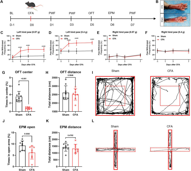
CFA inflammation in the left hind paw produces swelling and increased withdrawal to two von Frey forces over the following week, without the same significant change on the right. Open-field center and elevated-plus-maze open-arm time decrease, while total distance does not significantly change. The mice remain mobile but change where they explore.
Withdrawal and open-space exploration describe sensory responsiveness and anxiety-related behavior. Their co-occurrence prompts a search for shared central components: a population influencing both could help explain how inflammation affects more than local sensitivity.
This distinction matters: reduced overall movement could also reduce center time. Here, distance does not significantly change while open-area exploration decreases, allowing attention to the relationship between exploration choices and mechanical responses.
Source: Zhao Q et al. · Frontiers in neuroscience · CC BY
Original figure and English caption ↗
A cortical population becomes more responsive
After inflammation, ACC CRH/c-Fos colabeling increases, and CRH-labeled cells show stronger calcium responses to glutamate in slices. The population displays both activity-marker changes and altered input responsiveness. Direct manipulation then tests its behavioral contribution.
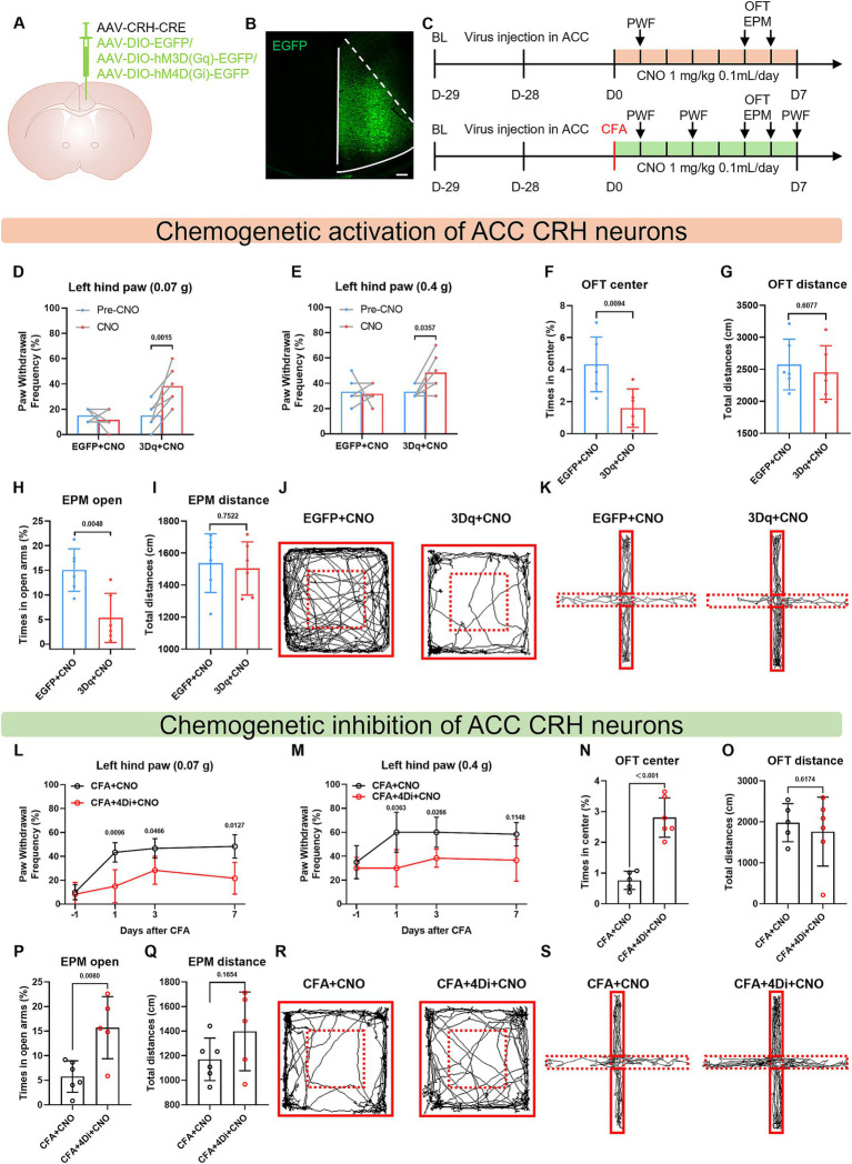
Repeated activation in healthy mice increases withdrawal and reduces open-area exploration. Inhibition in CFA mice shifts both measures oppositely, without significant distance changes. The day-seven comparison at 0.4 grams is nonsignificant. Population activity influences the phenotype without identical improvement at every time and force.
Source: Zhao Q et al. · Frontiers in neuroscience · CC BY
Original figure and English caption ↗
Signals from AM reach CRH-labeled cells
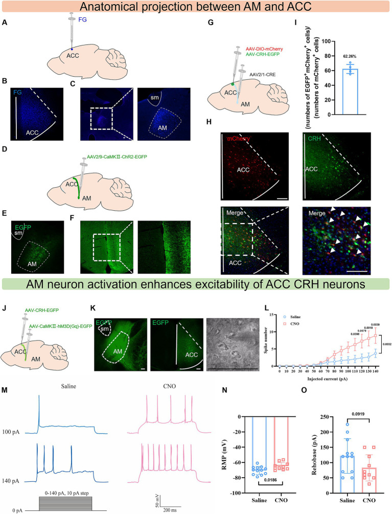
Where does input to these ACC cells originate? Forward and reverse tracing connects AM with ACC. Transsynaptic labeling finds CRH in approximately 62% of ACC cells receiving the labeled AM input. The thalamic route reaches a cortical population already implicated in behavior.
After AM activation in vivo, slice recordings show increased evoked firing and depolarized resting potential in CRH-labeled ACC cells, while rheobase changes are nonsignificant. Altering upstream activity changes downstream output readiness, linking anatomy with cellular responsiveness.
Source: Zhao Q et al. · Frontiers in neuroscience · CC BY
Original figure and English caption ↗
A memory-circuit node also contributes after inflammation
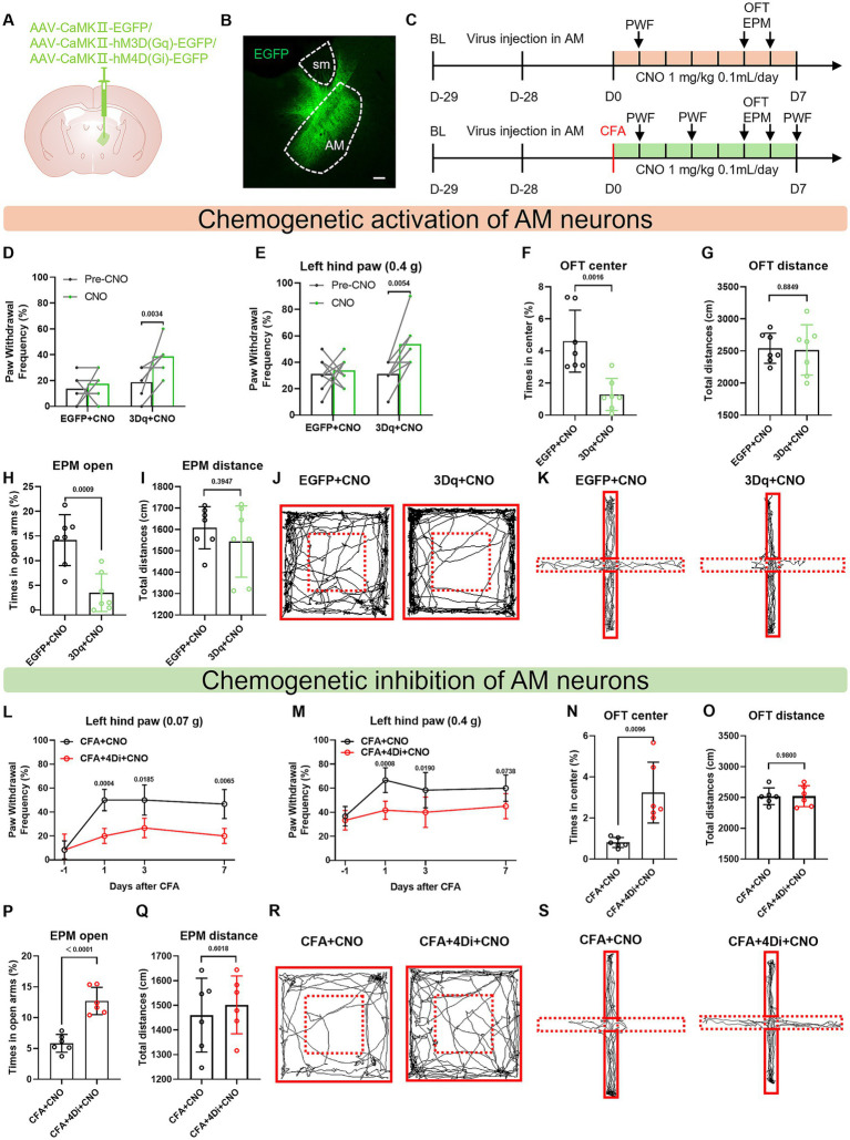
AM is familiar from the memory-related Papez circuit, yet it also changes after inflammation. CFA increases its slice calcium response to glutamate. Activating CaMKIIα-labeled AM cells increases withdrawal and avoidance in healthy mice; inhibition attenuates both in CFA mice. The day-seven 0.4-gram comparison remains nonsignificant.
Ablating this AM population before inflammation also attenuates later CFA-related changes. Prior cell removal and reversible activity manipulation provide complementary evidence about model development and behavior. A region’s familiar functional label does not exhaust its roles across bodily states.
Source: Zhao Q et al. · Frontiers in neuroscience · CC BY
Original figure and English caption ↗
Terminal manipulation changes behavior
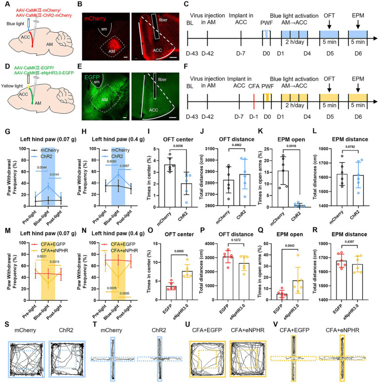
Manipulating AM terminals within ACC produces corresponding effects. Activation increases withdrawal in healthy mice and reduces open-area exploration after repeated intervention; inhibition improves these measures in CFA mice. Mechanical testing examines immediate light effects, while exploration follows multiday intervention. The cortical input has functional influence in both settings.
Source: Zhao Q et al. · Frontiers in neuroscience · CC BY
Original figure and English caption ↗
Do downstream cells matter during input activation?
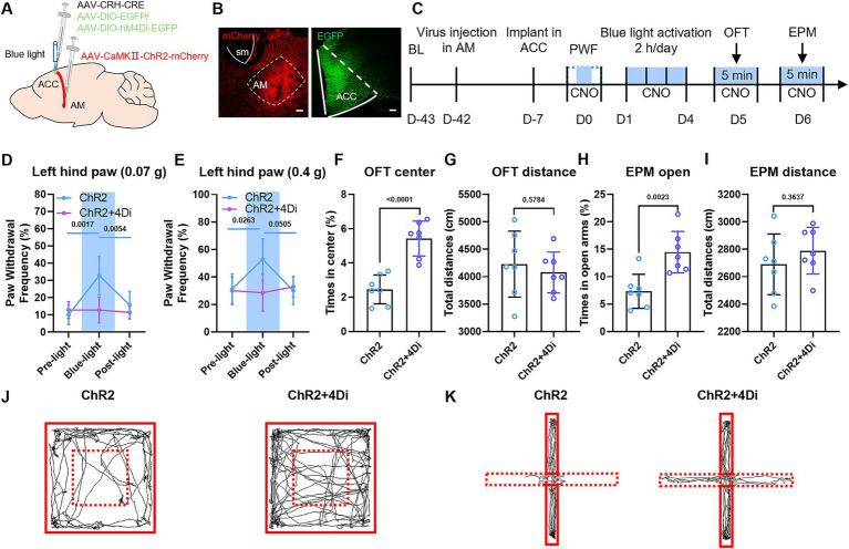
A final experiment combines upstream activation with downstream inhibition. Activating AM–ACC terminals while suppressing CRH-labeled ACC cells lowers withdrawal and increases center and open-arm time relative to input activation alone, without significant distance changes. Partial reversal of anxiety-related changes shows that this cortical population carries part of the input’s effect.
Source: Zhao Q et al. · Frontiers in neuroscience · CC BY
Original figure and English caption ↗
Two behaviors share part of a central route
In the male CFA model, AM input and CRH-labeled ACC cells connect mechanical sensitivity and open-area avoidance through a shared route. Persistent bodily disturbance relates not only to local sensory responses but also to thalamocortical organization. One input and its downstream population can influence both behavioral dimensions.