Pain and avoidance during intestinal inflammation: distinct roles of a brain connection
In a mouse colitis model, electroacupuncture accompanies reduced hypersensitivity and anxiety-like behavior. Hippocampal-to-prefrontal cells contribute differently to distinct pain responses.

Intestinal inflammation can be accompanied by changes beyond the abdomen. Hypersensitivity and avoidance of exposed spaces can emerge together in animal models. A 2025 Molecular Pain study follows the ventral hippocampus (vHPC)–medial prefrontal cortex (mPFC) connection to investigate how these changes meet. Electroacupuncture improves visceral responses and anxiety-like behavior, yet reactivating the projection-defined cells does not counteract every improvement. That distinction is central to understanding the brain contribution.
Inhibiting mPFC-projecting vHPC cells improves hypersensitivity and anxiety-like behavior in colitis-model mice. Activation partially counteracts electroacupuncture benefits for visceral responses and anxiety-like behavior without clearly reversing the hindpaw mechanical-threshold benefit.
Contents · 7 sections
Visceral sensitivity accompanies hindpaw sensitivity
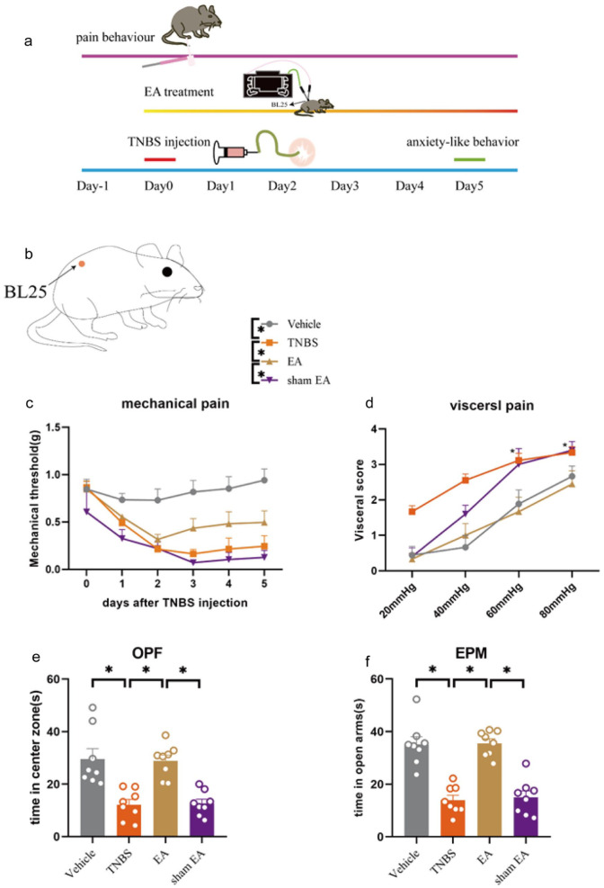
The study uses a TNBS-induced mouse colitis model. Compared with controls, model mice show stronger abdominal withdrawal responses to colorectal distension and lower hindpaw mechanical thresholds. Visceral and remote somatic sensitivity coexist, so the two nociceptive behaviors are followed separately.
The mice also spend less time in the open-field center and elevated-plus-maze open arms. These anxiety-like measures describe changes in choosing exposed versus relatively sheltered spaces. Their co-occurrence with hypersensitivity allows sensory responses and exploration to be compared in one model.
Source: Hu XF et al. · Molecular pain · CC BY-NC
Original figure and English caption ↗
Behavior improves as activity markers decline
Electroacupuncture-treated mice show higher hindpaw mechanical thresholds, lower colorectal-distension response scores and longer exposed-space time. Sham treatment does not produce comparable benefits. These behavioral changes occur together, raising the question of whether the same brain population contributes to each.
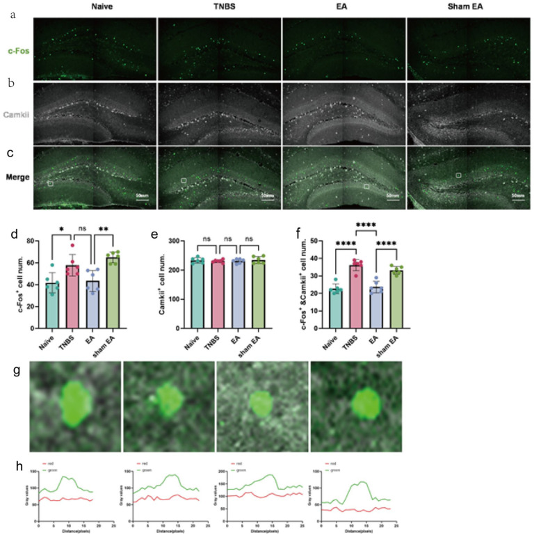
Overall excitatory-cell labeling does not clearly change in the tissue analysis; differences concentrate in the fraction also carrying c-Fos. The pattern therefore concerns activity state within the labeled population rather than an increase or decrease in its size.
Excitatory-cell c-Fos activity markers increase in both vHPC and mPFC in model mice and decline after electroacupuncture. Both areas change, but c-Fos summarizes activity over time. Calcium recordings next examine responses during specific behavioral events.
Source: Hu XF et al. · Molecular pain · CC BY-NC
Original figure and English caption ↗
Paw stimulation and entering exposed space accompany increased activity
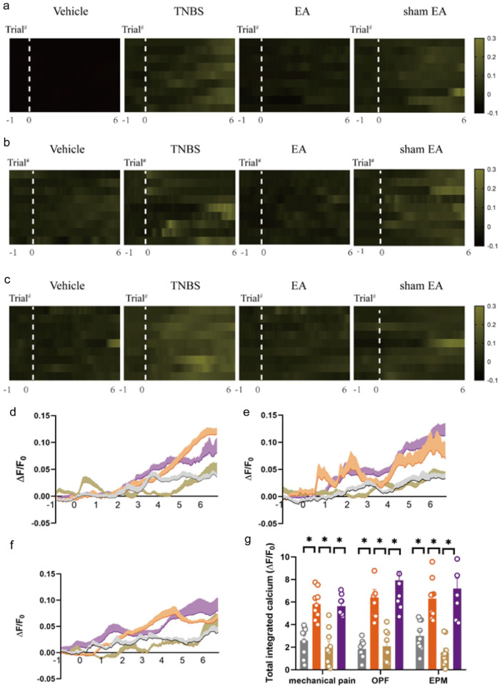
In model mice, vHPC excitatory cells show larger calcium responses to hindpaw mechanical stimulation and on entering the open-field center or open arms. After electroacupuncture, calcium signals associated with all three events diminish, matching the direction of behavioral improvement.
These recordings do not assign every cell one function; they show population activity in one region changing with different sensory and exploratory events. Following the events separately provides a clear comparison with prefrontal responses.
Source: Hu XF et al. · Molecular pain · CC BY-NC
Original figure and English caption ↗
Exploration responses decline while mechanical responses remain
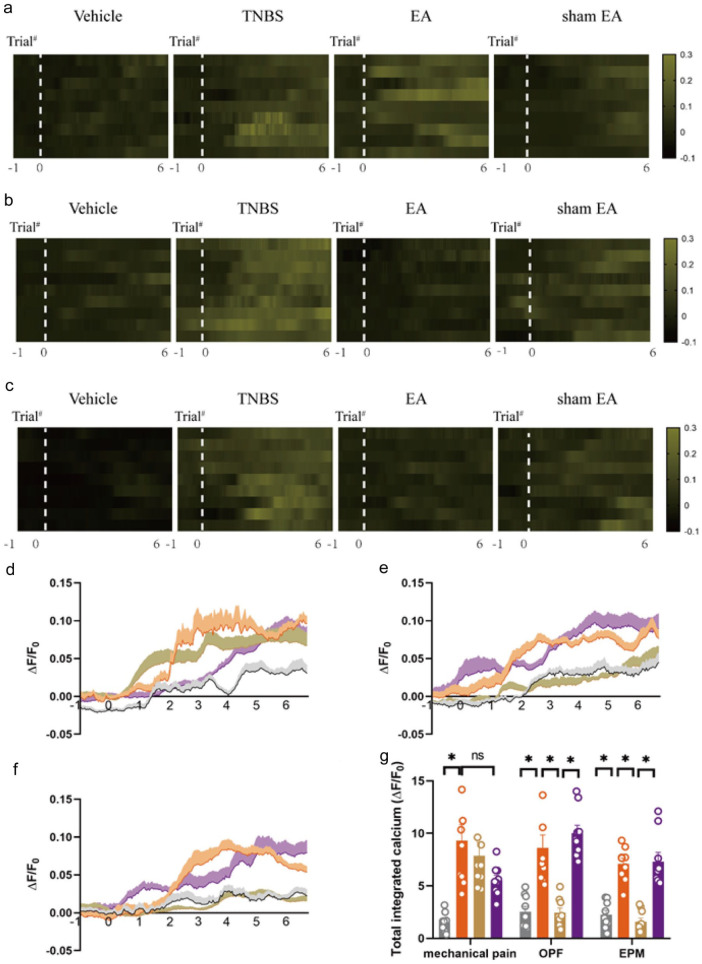
mPFC also shows larger event-related calcium responses in the model. After electroacupuncture, responses during center and open-arm events decline, while hindpaw mechanical responses show no clear change. The two areas therefore do not uniformly reduce every signal.
Improved mechanical sensitivity can coexist with a stable mechanical-response measure in this mPFC population. Sensory and emotion-related changes are connected but distinguishable. The study next intervenes in vHPC cells projecting to mPFC to test their behavioral contribution.
Source: Hu XF et al. · Molecular pain · CC BY-NC
Original figure and English caption ↗
Inhibition improves all three behavioral measures
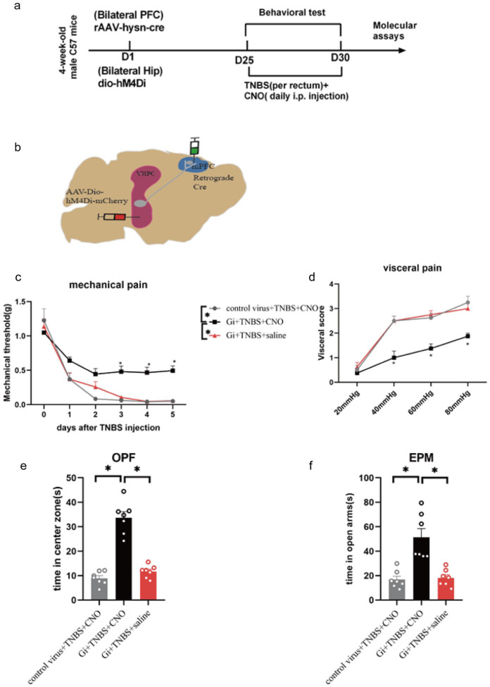
The study selects excitatory vHPC cells by their projection to mPFC and inhibits them. Model mice show higher hindpaw mechanical thresholds, lower abdominal withdrawal scores and longer time in the open-field center and open arms.
Reducing this population itself improves hypersensitivity and anxiety-like measures, strengthening the connection between brain activity and behavior. The next question is how much of the electroacupuncture benefit involves these cells.
Source: Hu XF et al. · Molecular pain · CC BY-NC
Original figure and English caption ↗
Visceral and exploration benefits recede without clear mechanical-threshold reversal
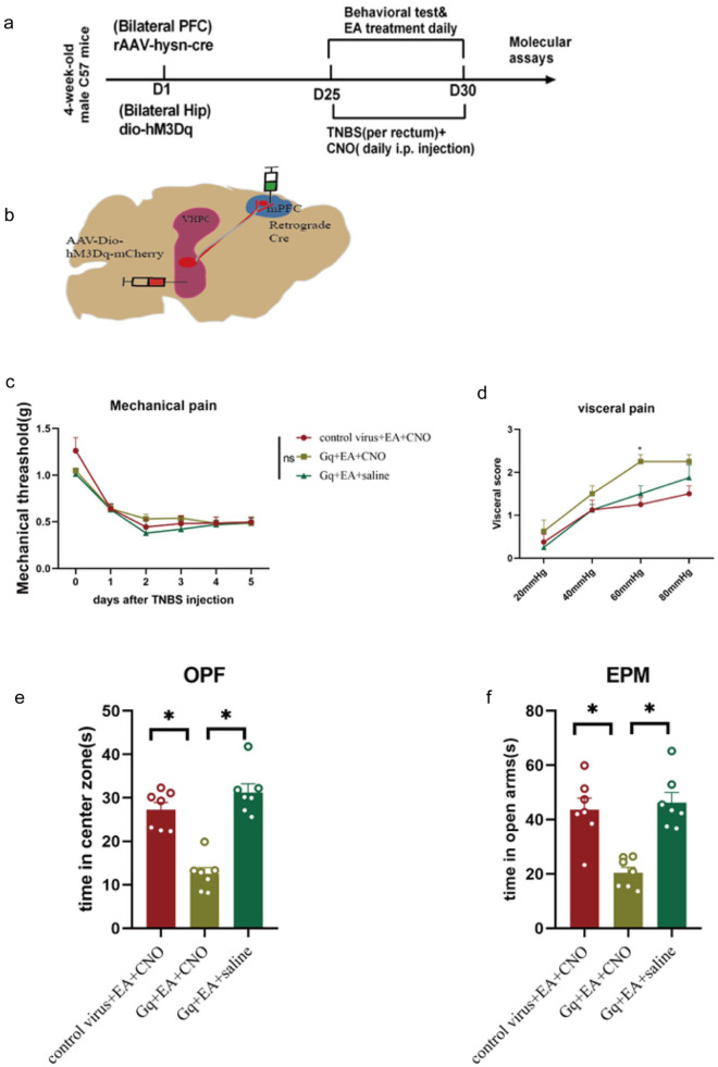
Reactivating the projection-defined cells in electroacupuncture-treated model mice increases visceral-response scores and reduces center/open-arm time, partially counteracting the benefits. Hindpaw mechanical thresholds, however, do not clearly differ among these groups, with no clear reversal of the mechanical benefit.
Inhibiting one population improves several measures, yet activating it counteracts only some electroacupuncture effects. The benefits thus involve multiple components: these hippocampal projection cells contribute closely to visceral and anxiety-like improvements, while mechanical improvement has a different pattern of involvement.
Source: Hu XF et al. · Molecular pain · CC BY-NC
Original figure and English caption ↗
Co-occurring changes can involve different routes
This adult male mouse study shows hypersensitivity and anxiety-like behavior changing together in a colitis model, then diverging during brain interventions. Focusing on prefrontal-projecting hippocampal cells explains part of the relationship between visceral responses and exploration and highlights that mechanical sensitivity and visceral responses are distinct processes.