Neurons and microglia reinforce nicotine-associated hypersensitivity
In male mice, prolonged nicotine exposure accompanies ACC neuronal hyperexcitability and microglial changes. Targeting either population or a candidate communication signal attenuates hypersensitivity.
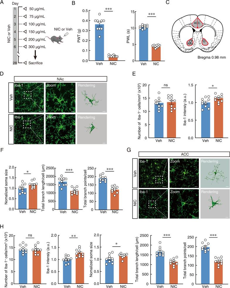
Why does the same light mechanical stimulus provoke stronger responses after prolonged nicotine exposure? A Journal of Neuroinflammation study published on March 21, 2023 finds two linked changes in the anterior cingulate cortex (ACC) of male mice: glutamatergic-associated neurons become more excitable, while microglial processes shorten and branch less. Interventions suggest reciprocal influences that may sustain elevated neuronal activity. CX3CL1–CX3CR1 signaling provides a candidate molecular link.
Reducing ACC glutamatergic-associated activity, pharmacologically targeting microglia or blocking CX3CR1 attenuates nicotine-associated mechanical and thermal hypersensitivity. Microglial morphological responses occur without significant increases in the classical inflammatory measures tested.
Contents · 6 sections
Microglial shape changes accompany hypersensitivity
Microglia are resident brain immune cells that also participate in network maintenance. Their branching processes reshape with changing conditions. After roughly four weeks of nicotine exposure, mice show lower mechanical withdrawal thresholds and shorter thermal withdrawal latencies, indicating increased responsiveness.
ACC Iba1-positive cell counts do not increase significantly, but somata enlarge, total process length falls and branching points decrease. The change therefore chiefly concerns cellular state rather than increased cell numbers. Similar morphology appears in the nucleus accumbens, examined as a comparison region, while subsequent interventions focus on ACC.
Source: Long DD et al. · Journal of neuroinflammation · CC BY
Original figure and English caption ↗
Morphology changes without matching inflammatory-marker increases
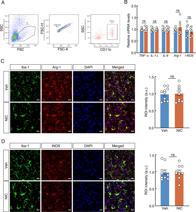
Does a reactive microglial shape mean that classical inflammation has broadly switched on? In isolated ACC microglia, TNF-α, IL-1β and IL-6 transcript levels do not differ significantly between groups. The additional tested markers Arg-1 and iNOS likewise show no significant changes.
Tissue labeling for the relevant markers gives similar results. The dissociation between morphology and these inflammatory measures suggests a distinct response profile, leaving other modes of communication as potential influences on neurons.
This dissociation reshapes the next question: how might microglia influence neuronal activity without an evident increase in the inflammatory factors tested? The investigation therefore moves beyond morphology to neuronal firing, sensory-event responses and changes in one population after intervening in the other.
Source: Long DD et al. · Journal of neuroinflammation · CC BY
Original figure and English caption ↗
Neurons cross the firing threshold more readily
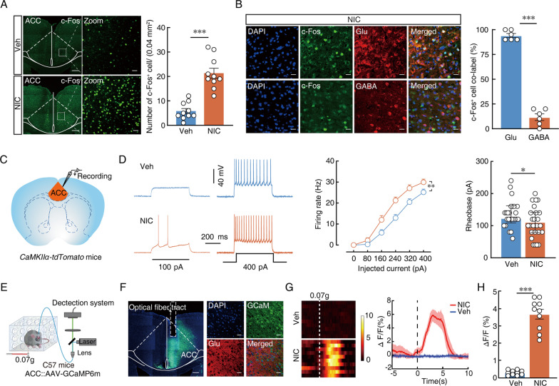
Meanwhile, more ACC cells express c-Fos, largely overlapping glutamatergic labeling. Slice recordings show more spikes for comparable input and a lower minimum current needed to trigger firing. The sampled neurons thus respond more readily to excitation.
Population calcium recordings connect the change to sensory events: light mechanical stimulation evokes larger signals in the nicotine group than in controls. Cellular excitability and stimulus-linked population activity provide complementary views of the region’s heightened responsiveness.
Source: Long DD et al. · Journal of neuroinflammation · CC BY
Original figure and English caption ↗
Reducing neuronal activity also changes microglia
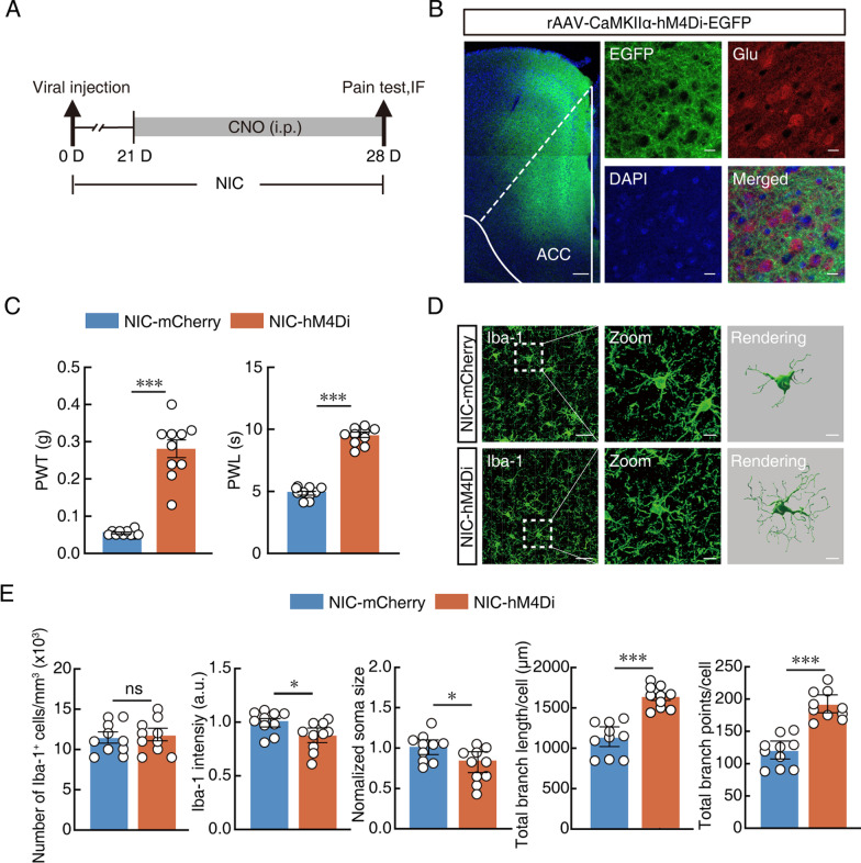
Coincident changes need not influence one another. Yet reducing ACC glutamatergic-associated neuronal activity raises mechanical thresholds and thermal withdrawal latencies, attenuating hypersensitivity. Microglial soma size, labeling intensity and branching also shift in the opposite direction.
Processes lengthen and branch points increase, without a significant change in marker-positive cell counts. This provides intervention evidence that neuronal activity can influence microglial state, while the precise sequence of changes requires more continuous observation.
Source: Long DD et al. · Journal of neuroinflammation · CC BY
Original figure and English caption ↗
Intervening from the microglial side also attenuates sensitivity
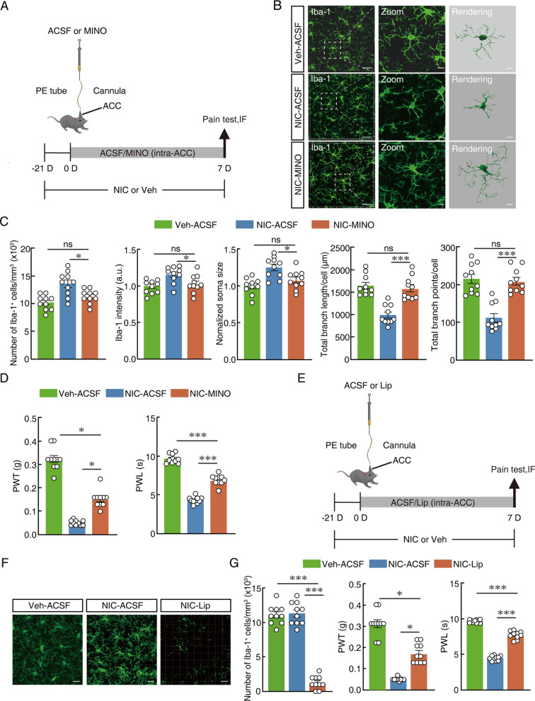
The study also intervenes from the microglial side. Local minocycline treatment attenuates morphological changes and mechanical and thermal hypersensitivity. Another pharmacological treatment that substantially reduces local Iba1-positive cells produces a similar behavioral direction.
Together, the treatments support microglial involvement in maintaining hypersensitivity. Improvements do not uniformly reach control levels, and the pharmacological interventions can affect other cellular processes. Combined with neuronal inhibition, the findings suggest a locally interacting network.
Source: Long DD et al. · Journal of neuroinflammation · CC BY
Original figure and English caption ↗
A candidate signal gives the interaction a molecular foothold
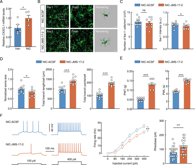
ACC tissue has higher CX3CL1 chemokine transcript levels. Blocking its receptor CX3CR1 reduces microglial soma size and labeling intensity, increases process length and branching, and attenuates hypersensitivity. In slices, sampled neurons fire less and require more current to initiate firing.
These results support a model in which neuron–microglia signaling helps sustain hyperexcitability. The evidence does not identify the CX3CL1 source cell by cell or directly measure synaptic pruning. In young adult male mice under prolonged nicotine exposure, the study identifies a local interaction: sustaining a hypersensitive network may involve altered microglia as well as spiking neurons.
Source: Long DD et al. · Journal of neuroinflammation · CC BY
Original figure and English caption ↗