How sensitivity spreads beyond a facial nerve injury
A male mouse study finds that thalamic VPM recruits body-sensory inputs after facial nerve injury. Early suppression delays distant sensitization, whereas later intervention has a different outcome.
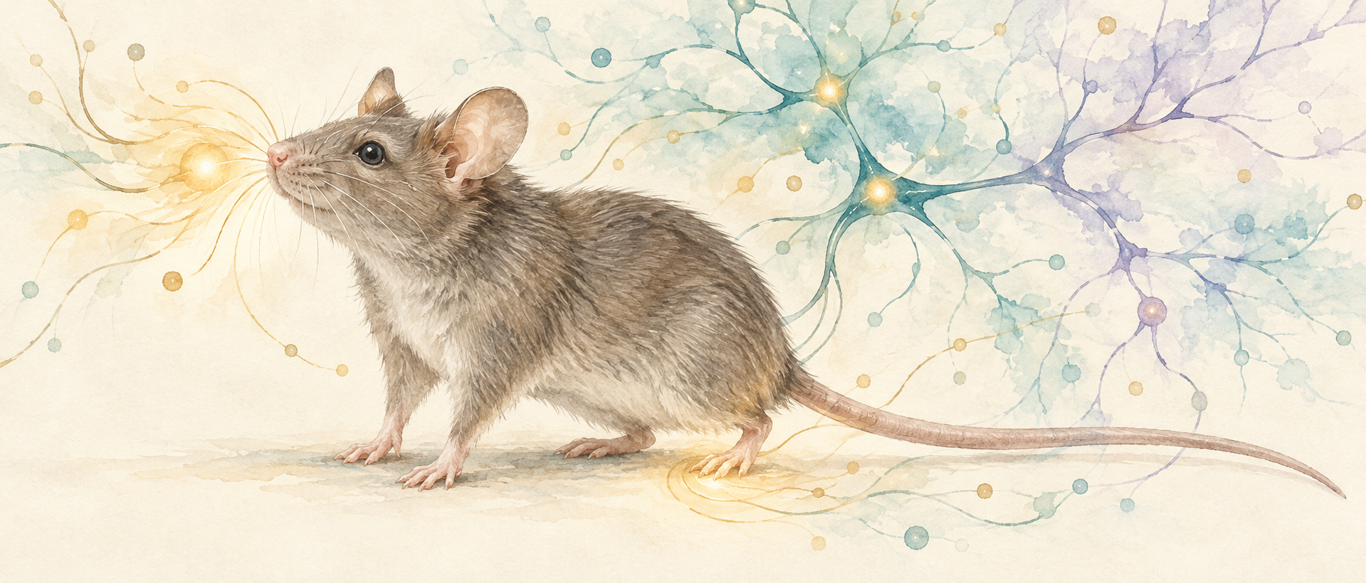
Why can a facial nerve injury make the hind paw and tail more sensitive? A study published in The Journal of Headache and Pain in 2024 follows changes in the ventral posteromedial thalamic nucleus (VPM). This relay, normally concerned mainly with facial sensation, becomes more active and recruits additional information from a body-sensory pathway after injury. The study also finds that the same recruited input contributes differently as sensitization spreads and after it is established.
After facial nerve injury, DCN-to-VPM input contributes to early development of distant sensitization. VPM and its posterior-insula output contribute to established sensitivity, with pathway suppression differing across early and later stages.
Contents · 7 sections
Sensitivity extends from the face to the body
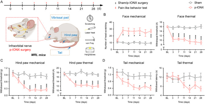
After partial infraorbital nerve injury, male mice scratch more in response to facial stimulation and have lower thermal response thresholds. Days later, hind-paw mechanical and thermal thresholds fall, with corresponding tail changes. An initially localized facial injury accompanies altered responses in distant body regions.
Facial and limb information normally travels mainly through different thalamic relays: VPM favors the face, while neighboring ventral posterolateral nucleus, VPL, favors the trunk and limbs. Distant sensitization raises the question of whether this division of sensory processing changes.
Source: Du Y et al. · The journal of headache and pain · CC BY-NC-ND
Original figure and English caption ↗
Changes first concentrate in VPM
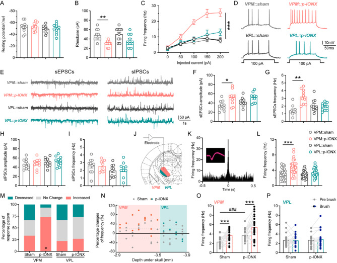
One week after injury, VPM neurons become easier to activate, fire more spontaneously, and receive stronger excitatory synaptic input. Corresponding VPL measures do not clearly change; VPM resting potential and inhibitory currents also remain similar. The entire sensory thalamus does not increase activity together.
Facial brushing recruits more VPM cells with increased responses and produces stronger average firing after injury. A sensory relay begins amplifying stimulation-related responses. The change occurs around the emergence of distant sensitization, making VPM a focus of further analysis.
Source: Du Y et al. · The journal of headache and pain · CC BY-NC-ND
Original figure and English caption ↗
A facial relay responds to distant stimulation
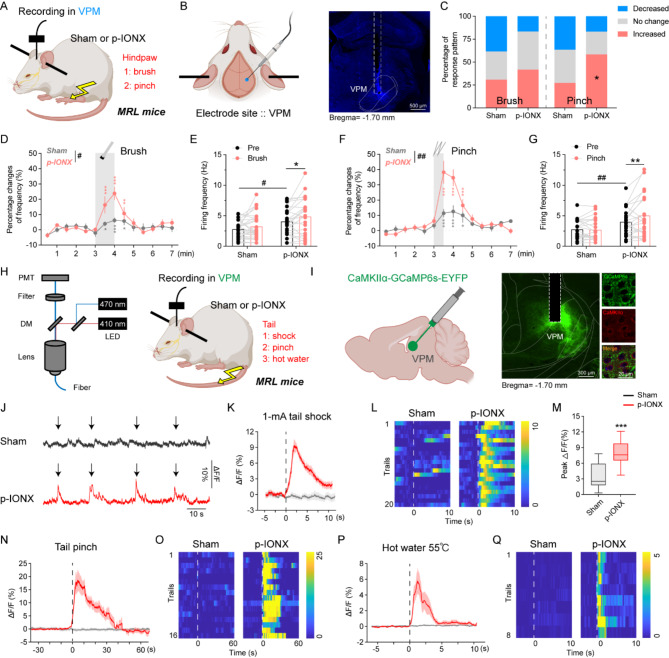
Hind-paw brushing or noxious stimulation evokes stronger VPM firing in injured mice than controls. The shift is not simply from no response to a response: controls can show mild responses, while injury produces stronger amplification. A relay biased toward facial information responds to distant stimulation.
Calcium recordings in awake mice likewise show clear VPM increases during different noxious tail stimuli after injury. Distant responses are thus observed in awake population activity as well as single-cell recordings under light anesthesia.
Source: Du Y et al. · The journal of headache and pain · CC BY-NC-ND
Original figure and English caption ↗
Lower relay activity eases sensitive responses
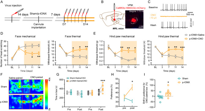
After sensitization is established, reducing VPM excitatory-cell activity eases facial and hind-paw mechanical and thermal sensitivity. Effects span two body regions, complementing the distant-stimulation recordings. VPM activity contributes to the measured sensitivity rather than merely accompanying it.
Mice also prefer the place paired with this inhibitory condition. In the pain model, that choice supports relief of sensitivity and associated aversion, extending the observations beyond stimulus-evoked responses.
Source: Du Y et al. · The journal of headache and pain · CC BY-NC-ND
Original figure and English caption ↗
The output reaches posterior insula
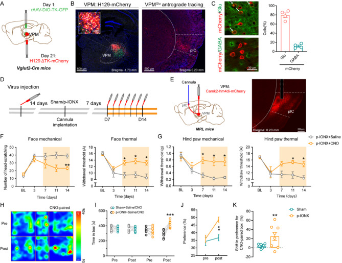
VPM projects to posterior insular cortex, pIC, where labeled downstream cells predominantly show excitatory characteristics. Reducing VPM terminal activity within pIC likewise eases facial and hind-paw sensitivity and produces preference for the corresponding place.
The findings connect activity in the relay with its cortical output. Information from different body regions can converge through VPM and influence sensitivity and aversion through a posterior-insula-associated pathway.
Source: Du Y et al. · The journal of headache and pain · CC BY-NC-ND
Original figure and English caption ↗
The relay recruits additional body-sensory input
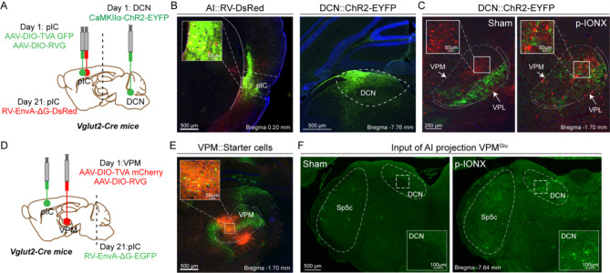
Tracing VPM input sources reveals an altered composition after injury: labeling proportions increase in body-related cortex and dorsal column nuclei, DCN, while secondary somatosensory cortical labeling decreases. DCN changes are particularly prominent; these nuclei normally relay trunk and limb information.
Further tracing shows DCN terminals mainly in VPL in controls, extending into VPM after injury and connecting with pIC-projecting VPM cells. In slices, terminal activity evokes excitatory currents in VPM, supporting functionally recruited input.
Source: Du Y et al. · The journal of headache and pain · CC BY-NC-ND
Original figure and English caption ↗
One pathway has different early and later effects
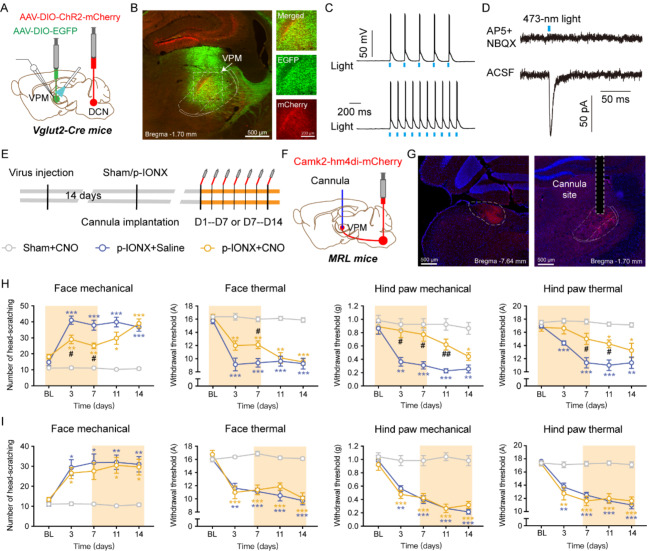
Reducing DCN-to-VPM terminal activity early after injury eases facial sensitivity and delays and mitigates hind-paw sensitization. After suppression ends, the effect fades and mechanical and thermal responses approach the control-intervention group. Early input modulation influences sensitization as it develops.
Once sensitization is established, suppressing the same DCN-to-VPM input does not clearly improve facial or hind-paw responses. This differs from effective suppression of VPM or its pIC output at that stage: recruited input contributes early, while established sensitivity depends differently on these circuit components.
These findings come from adult male mice with partial infraorbital nerve injury. They connect spreading sensitization with reorganized sensory circuitry and show that a pathway can contribute differently during development and persistence. Changing a relay node and changing a newly recruited input therefore produce different behavioral outcomes.
Source: Du Y et al. · The journal of headache and pain · CC BY-NC-ND
Original figure and English caption ↗