How does the thalamus respond to noxious input? Thirteen nuclei, different rhythms
A mouse thalamic map reveals differences within a shared response: nuclei vary in strength, timing and stimulus sensitivity, while PF and PO reach different networks.
A mechanical stimulus makes a mouse withdraw its paw while recruiting multiple brain regions. Do thalamic nuclei simply relay the same signal? Liu and colleagues compared population calcium activity across thirteen representative nuclei, revealing shared responses with different magnitudes and temporal profiles.
All thirteen nuclei respond to mechanical noxious stimulation. Classical medial pain-pathway nuclei show stronger, earlier responses overall, while PF and PO differ in timing and projection targets.
Contents · 6 sections
Looking inside the relay: thirteen distinct nuclei
The classical framework associates lateral pathways with sensory localization and discrimination, and medial pathways with affect, motivation and cognition. Understanding which regions respond earlier or for longer requires dynamic comparisons, previously complicated by differing animals, stimuli and recording conditions.
Using a common calcium indicator and comparable analysis, the team examined thirteen regions including AM, MD, PF and PO. The question expanded from whether a region responds to how strongly its signal rises and how it evolves within a broader thalamic map.
Shared recruitment, distinct waveforms
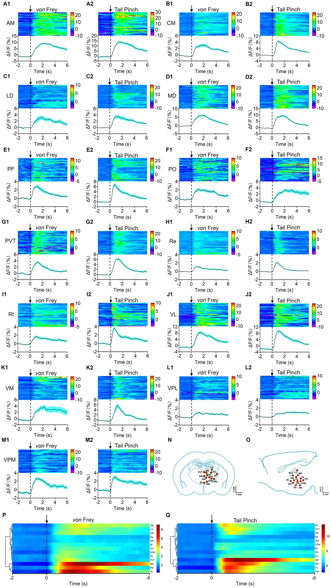
The team used a 2.0 g hind-paw von Frey stimulus and tail pinch as two mechanical noxious paradigms. The former reliably evoked withdrawal; the latter produced higher behavioral scores. Every recorded nucleus increased calcium signals under both conditions, extending recruitment beyond a few classical sensory nuclei.
Differences emerged within the shared response. AM occupied the highest signal cluster under both stimuli, while MD and VL remained relatively high; other nuclei changed clusters. Peaks, rise rates and persistence gave each response a distinctive profile.
Source: Liu YH et al. · CNS neuroscience & therapeutics · CC BY
Original figure and English caption ↗
A spatial pattern emerges: stronger, earlier medial responses
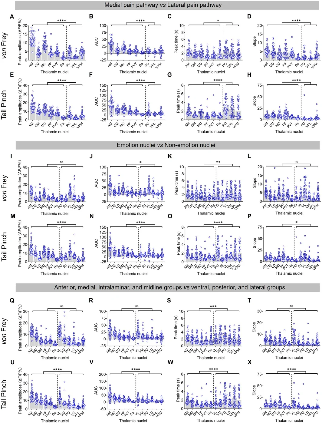
Trial-level clustering also supported a medial bias. When nuclei were grouped by classical medial and lateral pain pathways, the medial group showed higher peaks, larger areas under the curve, steeper rises and earlier peaks under both stimuli. This supports spatial organization of distributed responses.
Grouping added detail: affect-related or classical anatomical groupings differed significantly mainly under tail pinch, with no comparable differences under von Frey. Each framework organized a different set of nuclei, and stimulus conditions shaped when differences emerged.
Source: Liu YH et al. · CNS neuroscience & therapeutics · CC BY
Original figure and English caption ↗
Which responses change most between stimuli?
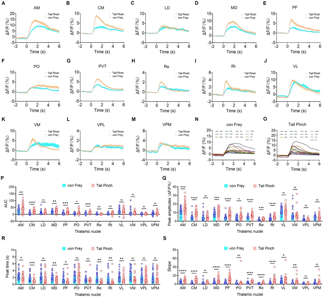
AM, CM, MD, PF, PO and PVT differed between stimuli in both peak and area. AM, CM, MD and PF also peaked earlier and rose more steeply under the stronger paradigm. Peak, area, time to peak and slope capture distinct aspects of a response.
VL, VM, VPL and VPM showed no significant peak or area differences, while other nuclei changed only some measures. The paradigms differed in site and form as well as intensity, and population recordings include movement and network feedback. The map describes responses under these concrete conditions.
Source: Liu YH et al. · CNS neuroscience & therapeutics · CC BY
Original figure and English caption ↗
PF and PO: different timing and output networks
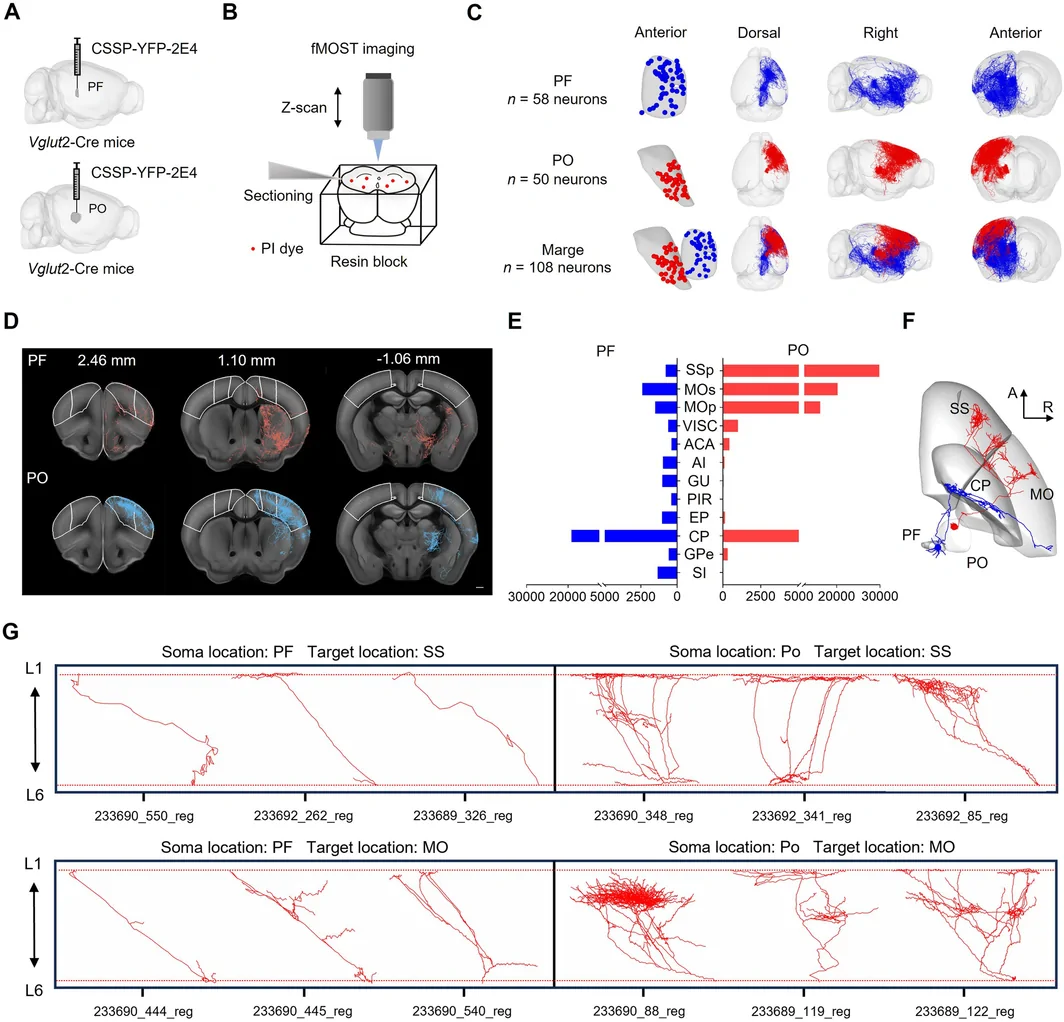
Among dorsal thalamic nuclei, PF had the shortest calcium time to peak and PO the longest. Reconstructions of 108 neurons from ten male Vglut2-Cre mice included 58 PF and 50 PO neurons. PF preferentially targeted striatal regions such as the caudoputamen, whereas PO more strongly targeted sensory and motor cortex.
Their cortical axons also differed: PF projections were relatively sparse and limited, while PO formed denser branches across layers. Striatal versus extensive sensorimotor cortical outputs provide context for their different rhythms and specific connections for future experiments.
Source: Liu YH et al. · CNS neuroscience & therapeutics · CC BY
Original figure and English caption ↗
A map that makes thalamic questions more precise
Which nuclei respond strongly, change with stimulus conditions, peak earlier and reach particular targets? Population activity and single-cell reconstruction turn a broad thalamic question into concrete comparisons and guide future tests of cells and connections.
The map comes from male mice under mechanical stimulation and uses population calcium signals. Cell subtypes, thermal stimuli, inflammation, movement and arousal components, and causal links between projection structure and timing remain promising directions.