How does electroacupuncture ease incision pain? Following a signal from brain to spinal cord
A mouse study finds reduced locus-coeruleus noradrenergic activity after incision; electroacupuncture re-engages this descending pathway and dampens exaggerated spinal responses to noxious stimuli.
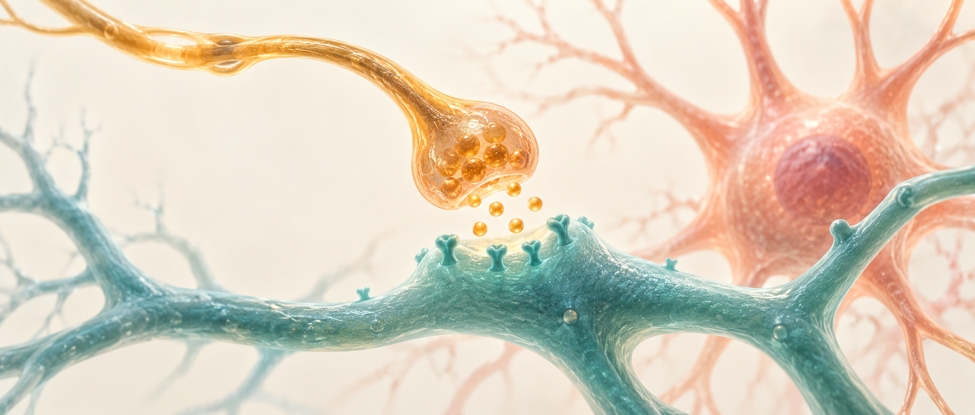
Pain arrives from injured tissue, but it does not simply travel upward unchanged. The brain also sends signals down to the spinal cord to adjust the strength of nociceptive transmission. A study published on May 19, 2025 in Advanced Science follows how electroacupuncture affects this volume control in a mouse plantar-incision model. After incision, noradrenergic cells in the brainstem locus coeruleus become less active and the corresponding spinal chemical signal falls. Electroacupuncture re-engages the descending pathway while reducing exaggerated spinal-cell responses.
Locus-coeruleus noradrenergic activity contributes importantly to electroacupuncture-induced relief of incision pain; spinal receptor and CaMKII-phosphorylation changes connect descending signaling to cellular responses.
Contents · 7 sections
Withdrawal changes—and so does contact with the ground
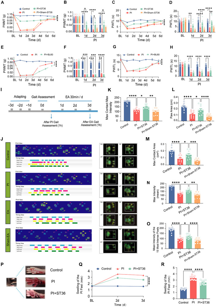
Following a plantar incision, mice become more sensitive to light mechanical stimulation and withdraw sooner from heat. These changes peak on the first day, persist for several days and then gradually recover. The first question is whether electroacupuncture alters this sensitive period. Stimulation at ST36 raises mechanical thresholds and heat-response latencies; sham stimulation does not produce the same improvement.
The changes also appear during walking. Incised mice reduce the area and intensity of ground contact with the injured paw; electroacupuncture improves these gait measures and reduces swelling. Stimulation at another tested site, BL60, does not clearly improve mechanical or heat sensitivity, indicating that the effect depends on the tested stimulation location.
Source: Chu WG et al. · Advanced science (Weinheim, Baden-Wurttemberg, Germany) · CC BY
Original figure and English caption ↗
A sensitized spinal cord accompanies a quieter locus coeruleus
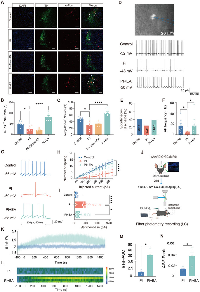
Electroacupuncture increases activity markers in several brain regions. The locus coeruleus attracts particular attention because its noradrenergic cells send modulatory signals to the spinal cord. After incision, their activity markers fall, fewer cells fire spontaneously and spontaneous firing slows. Cells without spontaneous activity also become harder to drive into repeated firing.
This direction challenges the intuition that pain excites every related cell. Electroacupuncture reverses these electrical changes, and recordings in living animals capture sustained increases in locus-coeruleus calcium signals during stimulation. Sensitivity at the injury coexists with low activity in a source of descending regulation; electroacupuncture may work partly by re-engaging that source.
Source: Chu WG et al. · Advanced science (Weinheim, Baden-Wurttemberg, Germany) · CC BY
Original figure and English caption ↗
Suppressing locus-coeruleus activity weakens relief
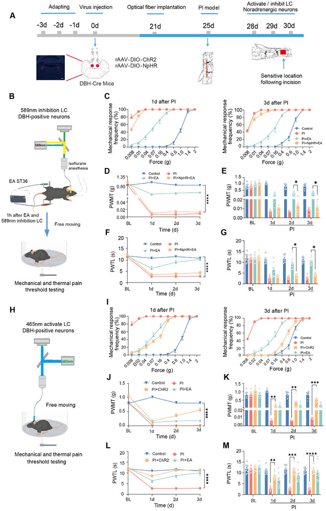
Seeing the locus coeruleus become active does not establish its contribution to altered pain responses. The study changes these cells’ activity directly. Suppressing them substantially weakens electroacupuncture-induced improvement in mechanical and heat sensitivity; activating them alone produces relief in the same direction in incised mice.
The opposite manipulations show that the locus coeruleus is more than a bystander. Activity in these cells supports the effect of electroacupuncture and can itself influence post-incision sensitivity. The next question becomes more concrete: does their signal reach the spinal dorsal horn, where nociceptive information is processed?
Source: Chu WG et al. · Advanced science (Weinheim, Baden-Wurttemberg, Germany) · CC BY
Original figure and English caption ↗
The change is local rather than a general rise in blood
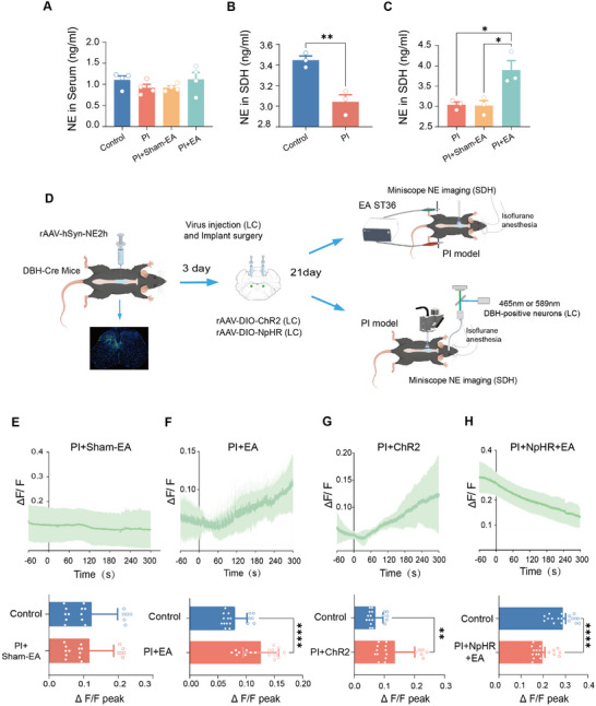
Noradrenaline levels in the spinal dorsal horn fall after incision and recover with electroacupuncture, while serum levels remain broadly unchanged. The change is therefore most apparent locally, where nociceptive information is processed, rather than as a general increase in circulating noradrenaline.
A molecular sensor in living animals makes the process visible: spinal noradrenaline signals increase during electroacupuncture, and activating locus-coeruleus cells produces a similar change. Suppressing those cells eliminates the electroacupuncture-induced spinal signal and can push it below baseline, directly supporting the link between brainstem activity and spinal chemistry.
Source: Chu WG et al. · Advanced science (Weinheim, Baden-Wurttemberg, Germany) · CC BY
Original figure and English caption ↗
The same noxious stimulus elicits a smaller response
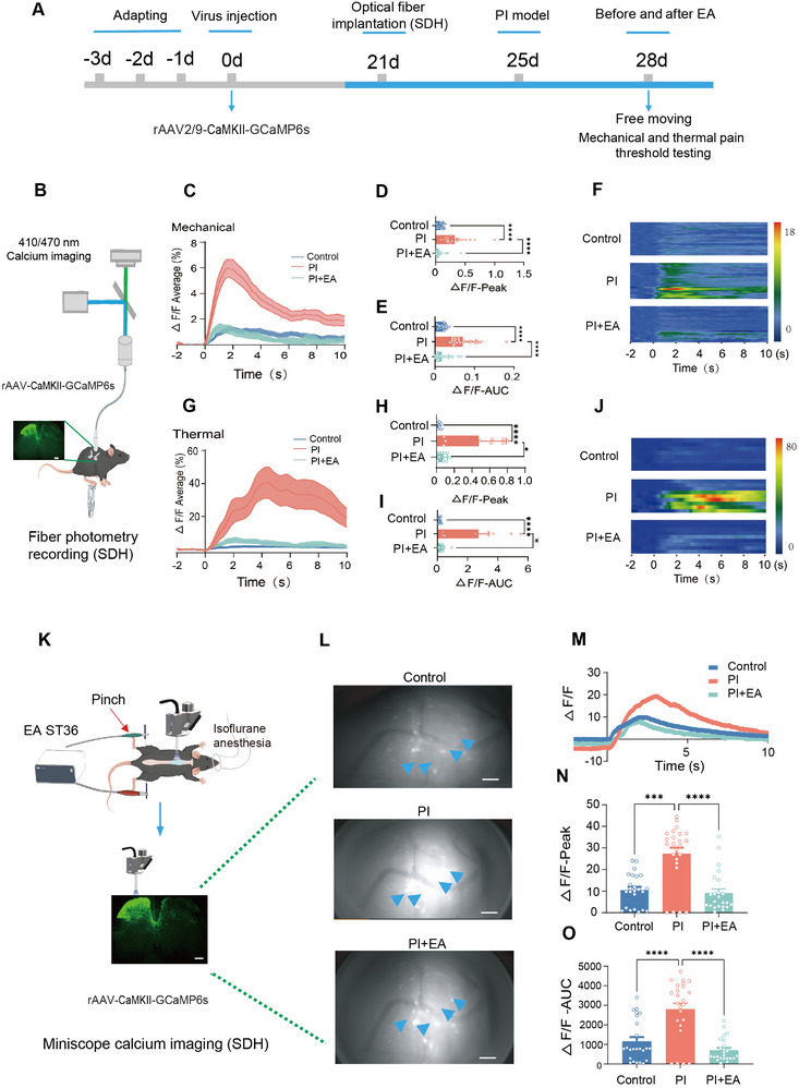
The spinal dorsal horn serves as an early processing station, receiving both nociceptive information from skin and regulation from the brain. After incision, mechanical and heat stimuli evoke stronger calcium responses in excitatory cells, showing sensitization within local processing as well as heightened sensitivity at the injured paw.
Electroacupuncture reduces these exaggerated population responses. Individual-cell recordings show a similar decrease in mechanically evoked responses. The findings connect withdrawal behavior to spinal-cell activity: relief accompanies reduced amplification of nociceptive information at an early processing stage.
Source: Chu WG et al. · Advanced science (Weinheim, Baden-Wurttemberg, Germany) · CC BY
Original figure and English caption ↗
Receptors and protein modification connect chemistry to response
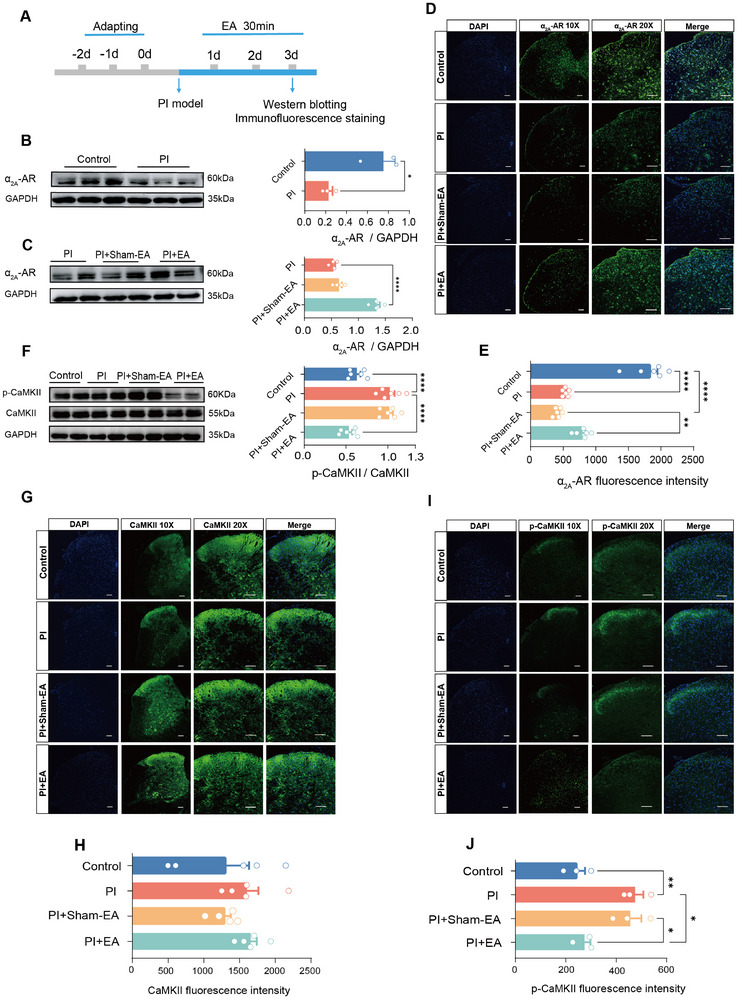
Noradrenaline acts through receptors. Spinal α2A-adrenoceptor expression falls after incision and rises again with electroacupuncture, while expression of three other tested receptor subtypes remains broadly stable. Total CaMKII, a protein involved in cellular plasticity, changes little, but its phosphorylation increases; electroacupuncture lowers that modification.
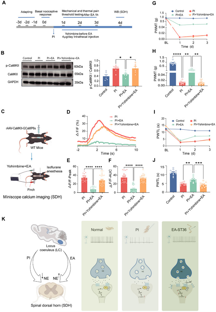
When yohimbine blocks spinal α2-receptor-related signaling, the improvements in CaMKII phosphorylation, cellular calcium responses and hypersensitivity are weakened. These results connect receptor signaling to changes at several levels and support involvement of α2A receptors and associated downstream regulation in the relief pathway.
Source: Chu WG et al. · Advanced science (Weinheim, Baden-Wurttemberg, Germany) · CC BY
Original figure and English caption ↗
Hypersensitivity can accompany weakened regulation
In a plantar-incision model including female and male mice, the study presents a two-direction story: spinal responses are amplified while noradrenergic activity in the locus coeruleus weakens. Electroacupuncture shifts both toward recovery, showing that pain sensitization can accompany reduced regulation as well as increased excitation.
Which intermediate steps carry peripheral stimulation to the locus coeruleus remains an open part of the pathway. The findings already connect stimulation near the skin to brainstem activity, spinal chemical signaling and cellular responses, revealing regulation that travels between brain and body.
Source: Chu WG et al. · Advanced science (Weinheim, Baden-Wurttemberg, Germany) · CC BY
Original figure and English caption ↗