Heat and pressure diverge along a lateral-habenula connection
In a mouse inflammation model, inhibiting the lateral-habenula connection to the rostromedial tegmental nucleus reduces thermal hypersensitivity without significantly improving mechanical thresholds.

An inflamed paw can become sensitive to both heat and pressure, yet the brain need not regulate both responses through identical routes. A study in The Journal of Headache and Pain focuses on the lateral habenula (LHb) and rostromedial tegmental nucleus (RMTg). Reducing this connection’s activity delays withdrawal from heat in mice without a comparable significant improvement in mechanical sensitivity. The contrast reveals a more specific sensory contribution of a node involved in processing negative information.
Changes in LHb–RMTg activity affect withdrawal from heat. In inflammation, inhibiting projection cells or their local terminals attenuates thermal hypersensitivity without significantly improving mechanical thresholds.
Contents · 6 sections
The same paw responds to distinct stimuli
Inflammation changes responses to noxious stimuli. The study measures time to paw withdrawal from heat and the force needed to evoke withdrawal with pressure. Shorter latency or lower force indicates greater responsiveness, but the readouts involve different sensory inputs.
Would altering one brain connection move both responses together? The researchers compare the readouts separately and find that they do not always move together. Their divergence becomes central to the story.
The lateral habenula sends information toward the brainstem
LHb participates in processing aversion, stress and negative outcomes. Its excitatory output reaches brainstem RMTg, a region rich in inhibitory neurons and connected with monoaminergic networks. This connection offers a route between sensory responses and broader brain states.
A brain region’s contribution depends on its inputs and outputs. LHb has more than one pain-related connection; this work examines a particular segment under different stimulus conditions. Comparing heat and pressure makes that contribution more specific.
The study focuses on RMTg-projecting LHb cells and their terminals. Chemogenetic experiments use male mice; terminal photoinhibition includes males and females. Manipulating these populations allows the two stimulus responses to be compared.
More activity, quicker withdrawal from heat
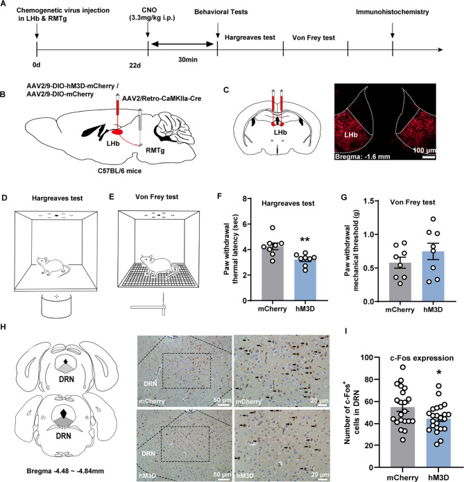
Activating RMTg-projecting LHb cells shortens mean heat-withdrawal latency from about 4.25 seconds in controls to 3.22 seconds. Mechanical withdrawal thresholds show no significant group difference. Thermal responses are the prominent change.
Inhibition produces a complementary result: heat-withdrawal latency increases, while mechanical-threshold comparisons remain nonsignificant. Opposite manipulations outline the connection’s contribution to thermal responses. Mechanical responses remain comparable in these tests.
Source: Sun Y et al. · The journal of headache and pain · CC BY
Original figure and English caption ↗
Thermal sensitivity remains adjustable after inflammation
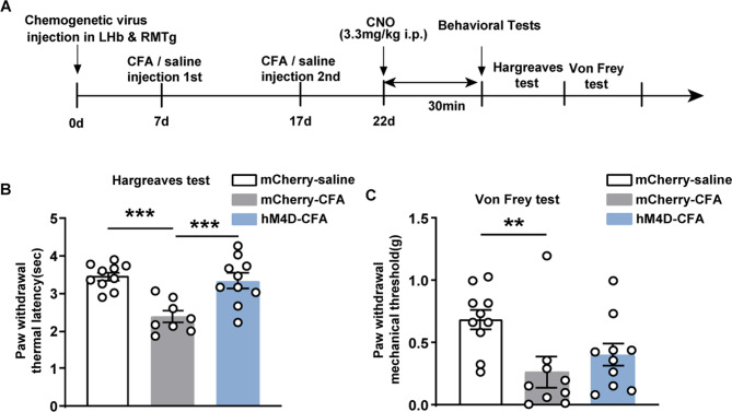
Inflammation produces both faster withdrawal from heat and lower mechanical thresholds. Inhibiting LHb projection cells raises thermal withdrawal latency from about 2.38 to 3.34 seconds, near the saline comparison group. Brain activity can influence behavioral responses accompanying peripheral inflammation.
Mechanical thresholds take a different course. Despite a numerically higher value in the inhibited group, the relevant comparison shows no significant improvement. The thermal change is accompanied by a stimulus-dependent difference in circuit contribution.
Source: Sun Y et al. · The journal of headache and pain · CC BY
Original figure and English caption ↗
The contrast persists at the terminals
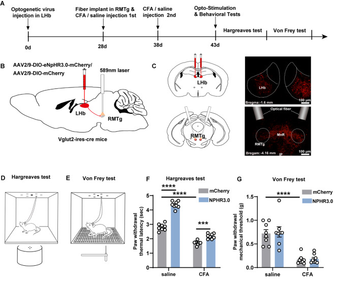
Photoinhibition targets terminals from LHb excitatory cells within RMTg. It lengthens thermal withdrawal latency in both saline and inflammatory conditions. Inflamed mice improve but remain below the noninflamed comparison level, indicating attenuated thermal hypersensitivity.
In this experiment, inflammatory controls have a thermal withdrawal latency of about 1.71 seconds, compared with 2.18 seconds under terminal inhibition. The change concerns a defensive response to stimulation; it does not itself establish resolution of inflammation. Brain modulation is examined while the peripheral inflammatory condition remains.
Mechanical responses again show no significant improvement. Projection-cell and local-terminal manipulations converge on the thermal–mechanical distinction, linking the effect more closely with LHb input reaching RMTg.
Source: Sun Y et al. · The journal of headache and pain · CC BY
Original figure and English caption ↗
The dorsal raphe provides a mechanistic clue
Activating LHb projection cells decreases c-Fos-positive cell counts in dorsal raphe (DRN), a region closely associated with serotonergic regulation. This motivates a possible LHb–RMTg influence on brainstem modulation of thermal sensitivity. The change in this brainstem node’s activity-related labeling provides a starting point for exploring downstream mechanisms.
The mouse study identifies a brain connection influencing thermal responses and reveals different dependencies for two forms of inflammatory sensitivity. As the body sends varied warning signals, brain modulation can produce different outcomes across sensory routes.