How do immune cells shape affect-related behavior after nerve injury?
A mouse study follows midbrain microglia, IL-1β signaling and a projection to the BNST to trace a brain pathway for behavior accompanying chronic pain.
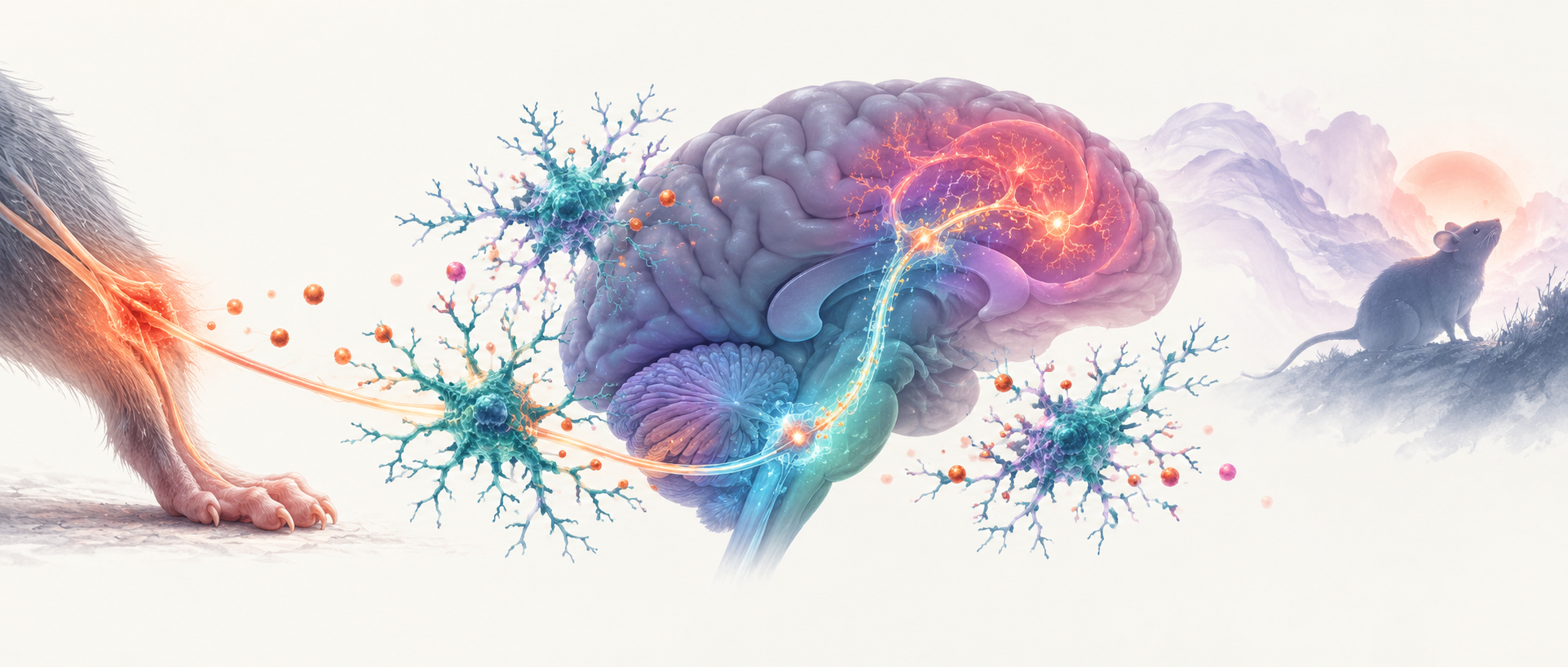
When pain persists, changes can extend beyond sensitivity to bodily stimuli into exploration and reward-related behavior. How do these changes arise in the brain? A 2026 Advanced Science study starts with peripheral nerve injury and turns to immune cells in the midbrain. In the adjacent ventrolateral periaqueductal gray and dorsal raphe region, or vlPAG/DRN, the researchers identify a chain from microglial inflammatory signaling to excitatory neurons and onward to the bed nucleus of the stria terminalis, or BNST. Molecular and projection-level findings reveal its influence on behavior accompanying pain.
In male mice, microglial NLRP3–IL-1β signaling connects vlPAG/DRN excitatory neurons with BNST GABAergic cells and modulates several negative-affect-related behaviors after nerve injury.
Contents · 7 sections
Exploration and reward change alongside pain
Mice in the spared nerve injury, or SNI, model developed persistent mechanical allodynia. By six weeks after injury, they also explored the open-field center less, remained immobile longer in tail-suspension and forced-swim tests, and showed lower sucrose preference. Different tasks captured several facets of altered engagement with the environment and reward after peripheral injury.
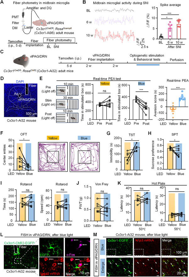
During this period, microglia in vlPAG/DRN showed changes in number and morphology, with elevated in vivo calcium activity at six and ten weeks after injury. As resident brain immune cells, microglia can sense and respond to their local environment. Their changes alongside behavior prompted a new question: can their activity help organize these behaviors?
Microglial activity changes behavior while sensory thresholds remain similar
Even without prior peripheral injury, increasing vlPAG/DRN microglial activity produced related changes. Mice avoided a stimulation-paired chamber, explored the open-field center less, remained immobile longer and showed lower sucrose preference. Optogenetic and chemogenetic strategies yielded convergent results, identifying a functional behavioral node rather than only a cellular marker accompanying injury.
Rotarod performance, mechanical thresholds and thermal sensitivity remained similar as these behaviors changed. Sensory sensitization and affect-related behavior therefore showed distinct patterns of modulation. Nearby VGLUT2-positive neurons had increased c-Fos labeling, while microglia expressed Nlrp3. Together, these clues pointed toward a connection between inflammatory molecules and local excitatory neurons.
Source: Yang J et al. · Advanced science (Weinheim, Baden-Wurttemberg, Germany) · CC BY
Original figure and English caption ↗
NLRP3 links the immune response with behavioral change
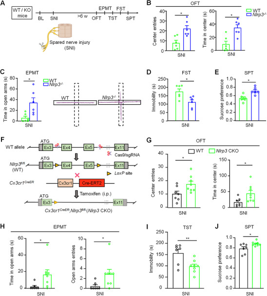
The NLRP3 inflammasome contributes to IL-1β maturation, offering a molecular route by which immune cells can influence other cells. After nerve injury, Nlrp3 and related inflammatory transcripts increased in vlPAG/DRN. Global Nlrp3 deletion attenuated negative-affect-related phenotypes, and conditional deletion in microglia improved exploration and other behavioral measures.
The molecular change did not appreciably impair baseline physiology or behavior, and improvement accompanied preserved sensory thresholds and motor measures. The work thus gained evidence at both the cell-type and molecular levels. The next step moved to the local midbrain network: which cells receive the inflammatory signal, and how does that reception change neuronal activity?
Source: Yang J et al. · Advanced science (Weinheim, Baden-Wurttemberg, Germany) · CC BY
Original figure and English caption ↗
IL-1β recruits local excitatory neurons
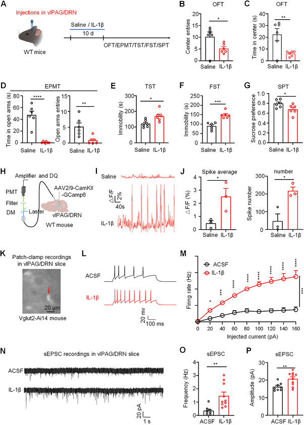
Local IL-1β in naïve mice changed exploration, immobility and reward preference while increasing vlPAG/DRN neuronal calcium activity. VGLUT2-positive cells in slices also showed greater firing and excitatory synaptic input. An immune-system molecular signal was thus connected to neuronal electrical activity.
These excitatory cells co-expressed Il1r1 and Vglut2, placing them in a position to sense IL-1β. Reducing their Il1r1 expression or blocking IL-1R1 locally attenuated the corresponding post-SNI behavioral deficits. Suppressing this neuronal population also improved behavior, making these signal recipients a functional link between local inflammation and behavioral change.
Source: Yang J et al. · Advanced science (Weinheim, Baden-Wurttemberg, Germany) · CC BY
Original figure and English caption ↗
The midbrain signal reaches the BNST
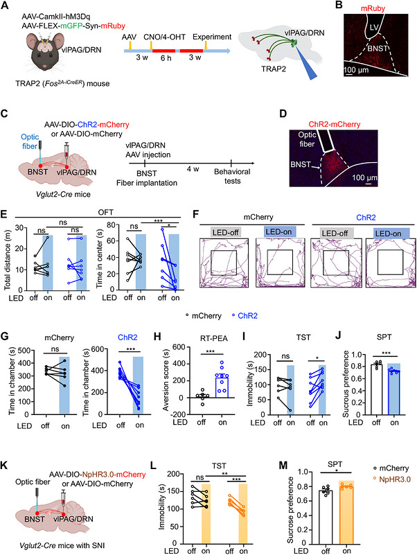
Where does the signal travel after recruiting these neurons? Circuit tracing pointed to the BNST, a region involved in sustained threat and affective regulation that receives excitatory projections from vlPAG/DRN. Activating the projection’s BNST terminals reduced center exploration, produced real-time place avoidance, increased immobility and lowered sucrose preference, with overall locomotion remaining similar.
In mice that had undergone SNI, suppressing the same projection reduced tail-suspension immobility and restored sucrose preference. Activation and inhibition connected this projection with defined behavioral states, establishing a network segment that links local inflammatory influences to a more distant behavioral regulatory node.
Source: Yang J et al. · Advanced science (Weinheim, Baden-Wurttemberg, Germany) · CC BY
Original figure and English caption ↗
Excitatory input recruits BNST GABAergic cells
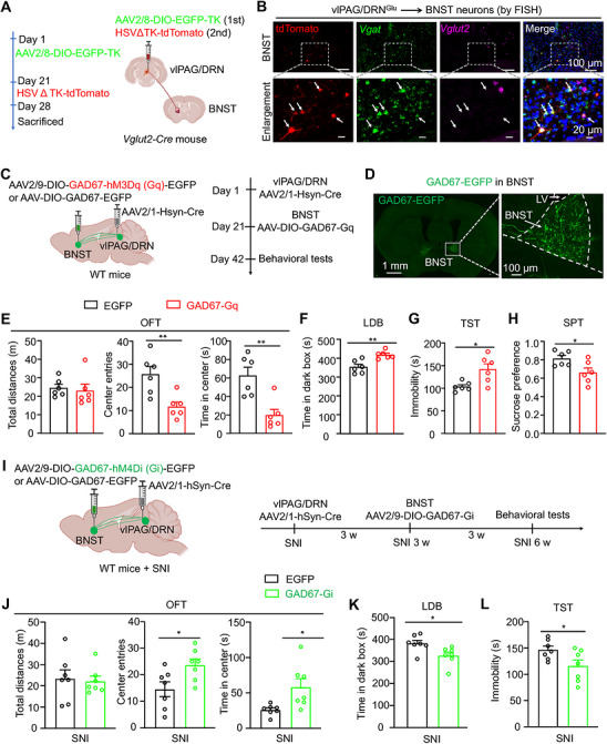
Which BNST cells receive the input? Trans-synaptic labeling showed that vlPAG/DRN recipients predominantly co-expressed Vgat, identifying GABAergic cells. Activating this input-receiving population reproduced related behaviors. Suppressing it under SNI improved center exploration, light–dark behavior and tail-suspension immobility, defining the projection’s endpoint by both cell identity and function.
The connection illustrates the layers of network regulation: excitatory input recruits an inhibitory cell population downstream. Behavioral outcomes depend on how these cells connect with the rest of the network. Following BNST outputs will help explain how immune-driven signals organize exploration, avoidance and reward responses across broader brain networks.
Source: Yang J et al. · Advanced science (Weinheim, Baden-Wurttemberg, Germany) · CC BY
Original figure and English caption ↗
Connecting pain-associated behavior to a neuroimmune network
From microglia to inflammatory molecules, and from local neurons to a BNST projection, the study connects peripheral injury with brain-based behavioral regulation across several levels. In multiple experiments, exploration or reward behavior changed while sensory thresholds and motor performance remained similar. This invites study of both how the body senses stimuli and how the brain changes behavior against a background of persistent pain.
The findings come from male mice, with local interventions spanning adjacent vlPAG and DRN. Sex differences, the individual contributions of the two regions, and the upstream recruitment of midbrain immune cells remain directions for future work. The identified chain offers a concrete starting point for studying immune state and neural circuitry as connected processes.