Communication between cingulate cortices increases pain sensitivity in mice
Excitatory connections link the left and right anterior cingulate cortices. Activating them increases mechanical and heat sensitivity; inhibition reduces some mechanical hypersensitivity after nerve injury, with effects that depend on body side and time.
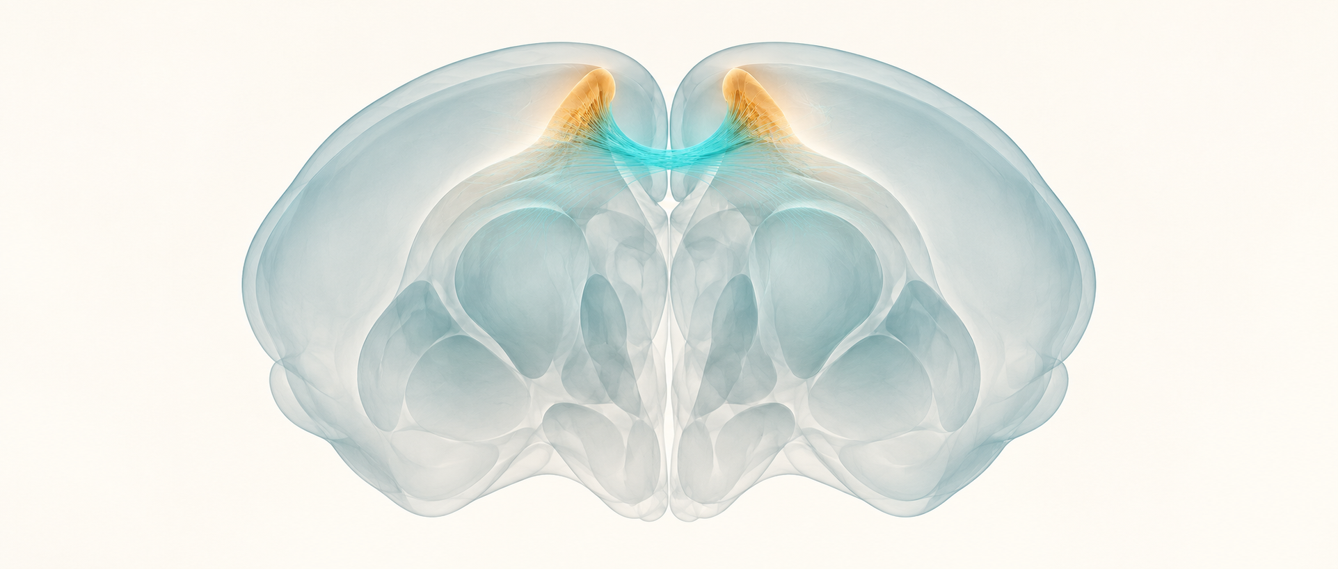
An injury to one hind paw can be followed by sensitivity on both sides of the body. How does pain processing cross the brain’s midline? A study published in Communications Biology on December 9, 2023 examines the connection between the two anterior cingulate cortices. Following fibers, synaptic currents and responses to touch and heat in adult mice, the researchers find that this cortical connection can facilitate pain-related responses.
The two ACCs communicate through glutamatergic excitation involving AMPA and, in some cells, kainate receptors. Activation increases mechanical and heat sensitivity. Inhibition improves mechanical sensitivity on the injured side across several post-injury time points, whereas its effect on the opposite paw is more time-dependent.
Contents · 7 sections
A working connection between the two ACCs
The anterior cingulate cortex (ACC) contributes to pain, emotion and action selection. Previous work found bilateral ACC changes after injury to one side of the body, but explaining communication requires examining the connection itself. Activity on both sides alone does not show that one side drives the other.
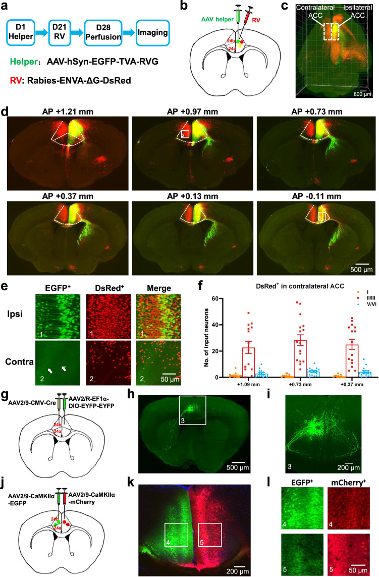
Tracing reveals fibers crossing the corpus callosum between the ACCs, with connected neurons in both superficial and deep layers. Labeled fibers from one side appear among cells on the other, providing an anatomical basis for bilateral communication.
Source: Li XH et al. · Communications biology · CC BY
Original figure and English caption ↗
Glutamate makes the connection excitatory
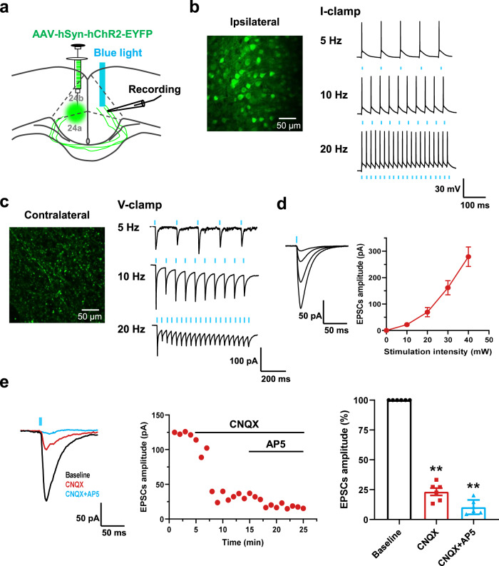
Does the connection transmit signals? Stimulating fibers from one ACC evokes excitatory synaptic currents in contralateral pyramidal cells, including responses to repeated stimulation. Glutamate-receptor blockade greatly reduces these currents. Further recordings identify a major AMPA-receptor component and an additional kainate-receptor component in some cells.
Input strength also matters for calcium signaling. Weaker inputs that do not trigger spikes produce no significant increase in the recorded somatic calcium signal. Inputs that evoke action potentials, especially bursts, produce larger calcium responses. Communication across the midline can thus alter both electrical activity and intracellular signaling.
Source: Li XH et al. · Communications biology · CC BY
Original figure and English caption ↗
Stronger communication changes withdrawal responses
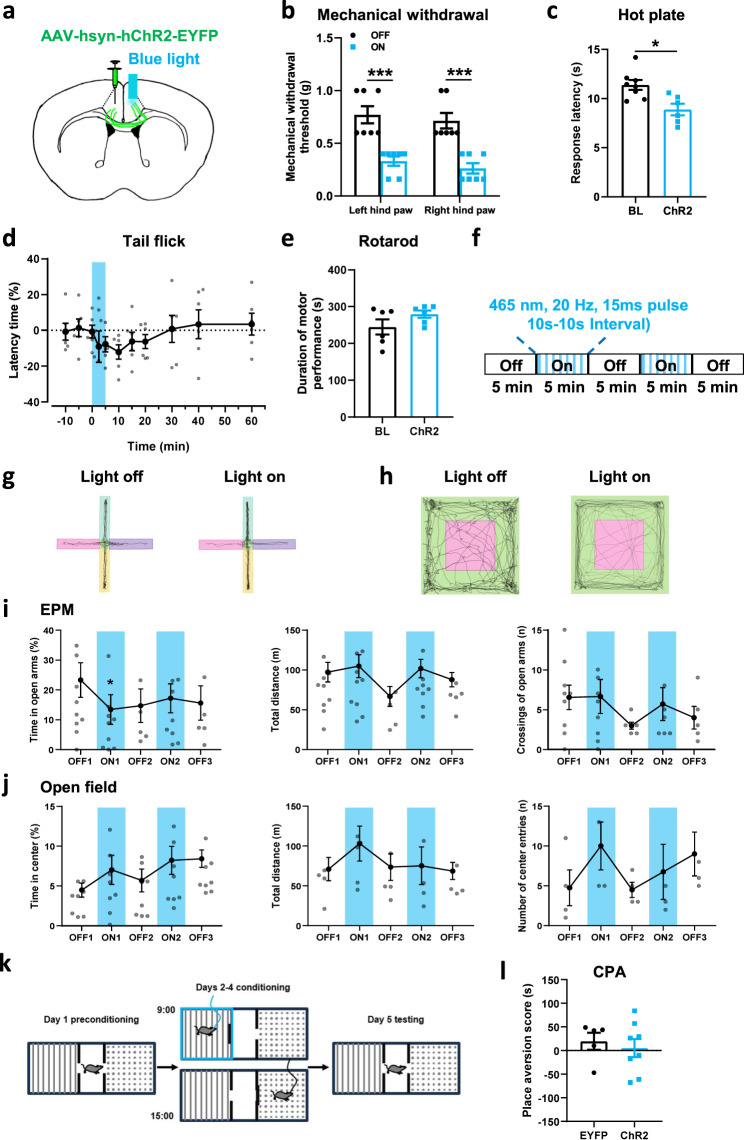
In uninjured male mice, activating the connection lowers mechanical withdrawal thresholds in both hind paws and shortens hot-plate and tail-flick response latencies. Rotarod performance does not change significantly, indicating no evident accompanying change in motor performance in that assay.
Affect-related measures do not change uniformly. Time in the open arms of the elevated plus maze decreases, but entry counts do not change significantly. Open-field center exploration and conditioned place aversion also show no significant differences. Heightened stimulus sensitivity and learned avoidance of a location therefore have different outcomes here.
Source: Li XH et al. · Communications biology · CC BY
Original figure and English caption ↗
Females also become more sensitive
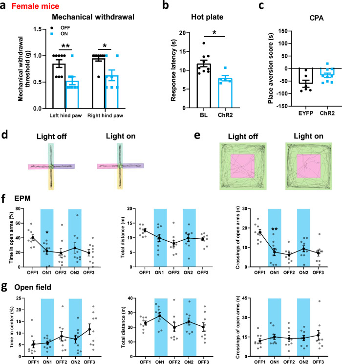
Females also show lower mechanical thresholds in both hind paws and faster hot-plate responses, extending the sensory finding beyond males. In the elevated plus maze, both open-arm time and entries decrease in females, whereas the significant male change is in time spent there.
Open-field center exploration and place-aversion scores remain without significant changes in females. Increased mechanical and heat responsiveness is shared across the cohorts, while the exploration measures vary with assay and sex.
Source: Li XH et al. · Communications biology · CC BY
Original figure and English caption ↗
The whole region has a broader reach
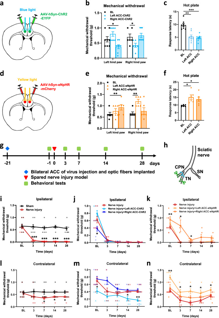
The researchers also manipulate the activity of an entire unilateral ACC population. In uninjured mice, activation lowers the opposite paw’s mechanical threshold, whereas inhibition raises it. Hot-plate responses become faster with activation and slower with inhibition, demonstrating a capacity of unilateral ACC activity to modulate nociceptive responses.
The ACC also connects to several subcortical targets. When interhemispheric fibers are inhibited, however, only one paw’s mechanical threshold increases significantly in uninjured animals; the other paw and hot-plate latency show no significant change. The behavioral reach of the entire ACC and its interhemispheric connection is therefore not identical.
Source: Li XH et al. · Communications biology · CC BY
Original figure and English caption ↗
Inhibition has different time courses in the two paws
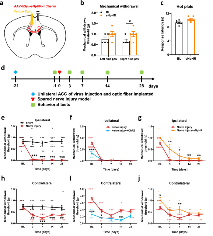
After unilateral peripheral nerve injury, both the injured-side and opposite hind paws become mechanically hypersensitive. Inhibiting the interhemispheric connection raises the injured paw’s threshold on days 3, 7, 14 and 28. A significant increase in the opposite paw occurs only on day 7. The connection contributes to bilateral sensitivity with different temporal patterns.
Conversely, activating the connection does not significantly lower the injured paw’s threshold further, but increases opposite-paw sensitivity at some time points. In the injured network, increasing and decreasing activity in the same pathway do not always yield mirror-image outcomes.
Source: Li XH et al. · Communications biology · CC BY
Original figure and English caption ↗
Pain facilitation can involve communication between hemispheres
The study links a visible interhemispheric connection to excitatory transmission and pain-related behavior. Bilateral ACC changes gain an additional explanation: the two sides can influence stimulus responses through their connection as well as receiving inputs of their own.
The evidence comes from adult mice, using withdrawal, heat-response and exploration measures. Activating projection terminals may also recruit their parent cells and other branches. The findings reveal an interhemispheric contribution to pain facilitation and raise a specific question: how does injury reshape cooperation between the two ACCs and their downstream networks to produce different time courses on the two sides of the body?