How are pain sensation and negative affect separated in the paraventricular thalamus?
A mouse study dissects two anterior paraventricular thalamus projections, linking the bed nucleus pathway to mechanical hypersensitivity and the nucleus accumbens pathway to pain aversion.
Pain encompasses both a sensory-discriminative component (such as mechanical hypersensitivity) and an affective-motivational component (such as place avoidance and aversion). Although the anterior paraventricular thalamus (PVA) is a key midline thalamic hub implicated in nociceptive processing, whether mechanical hypersensitivity and pain-related aversion are co-encoded by overlapping PVA neurons or segregated across distinct neuronal ensembles and downstream pathways has remained unresolved. Published in Cell Reports on November 4, 2024, a study led by Dr. Chien-Chang Chen, Dr. Shi-Bing Yang, and Dr. Chi-Li Gong at Academia Sinica ("Separate anterior paraventricular thalamus projections differentially regulate sensory and affective aspects of pain") combined longitudinal miniscope single-cell calcium imaging, bidirectional optogenetics, viral circuit tracing, and simultaneous ChrimsonR stimulation with GCaMP6f miniscope imaging in mice to demonstrate that discrete PVA VgluT2+ glutamatergic ensembles and projections separately govern mechanical hypersensitivity (PVA VgluT2+→BNST) and pain-associated place avoidance (PVA VgluT2+→NAc).
Formalin-induced inflammatory pain recruits two non-overlapping ensembles of PVA VgluT2+ neurons (only 1% overlap): the PVA VgluT2+→BNST pathway induces persistent BNST neuronal hyperactivity to drive mechanical hypersensitivity lasting up to 3 days, whereas the PVA VgluT2+→NAc pathway selectively mediates pain-related real-time and conditioned place avoidance.
Contents · 6 sections
Intraplantar Formalin Persistently Recruits PVA VgluT2+ Neurons and Elevates In Vivo Single-Cell Calcium Activity
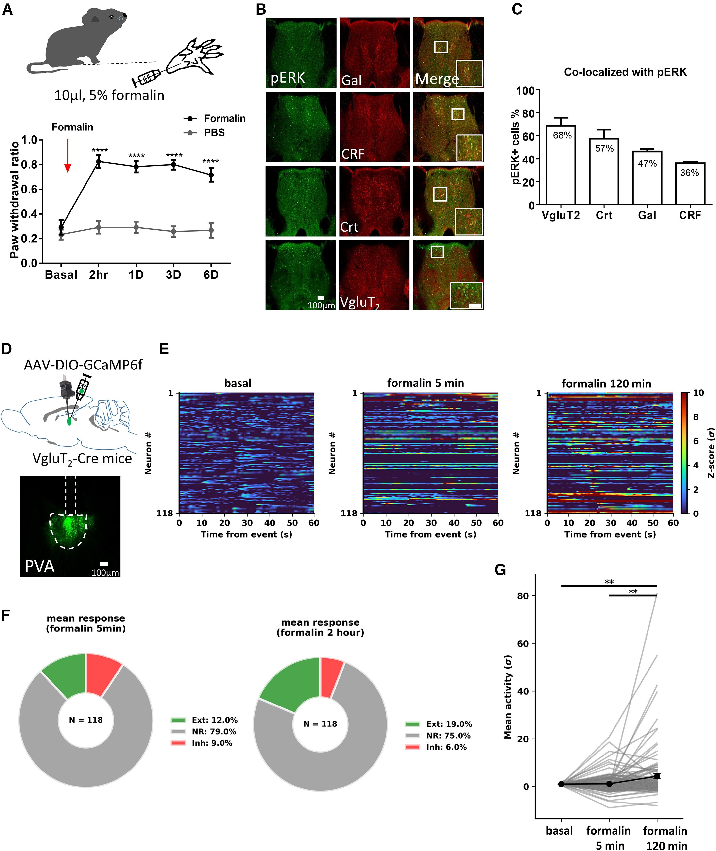
Intraplantar injection of formalin (10 μL, 5%) in mice significantly increased the mechanical paw withdrawal ratio (PWR) at 2 hours, 1 day, 3 days, and 6 days post-injection (p < 0.0001, n = 6). At 3 days post-formalin, phosphorylated ERK (pERK) colocalized with 68% of VgluT2+ glutamatergic neurons in the PVA, compared with 57% of calretinin+ (Crt+), 47% of galanin+ (Gal+), and 36% of CRF+ neurons.
Using a head-mounted miniature microscope (miniscope) to image GCaMP6f activity across 118 PVA VgluT2+ neurons in 5 freely moving VgluT2-ires-Cre mice, the authors found that the proportion of excited (Ext) neurons rose from 12.0% at 5 min post-formalin (79.0% non-responsive [NR], 9.0% inhibited [Inh]) to 19.0% at 2 hours post-formalin (75.0% NR, 6.0% Inh), accompanied by a significant elevation in mean Ca2+ activity at 120 min (p < 0.01).
Source: Selomon Assefa Mindaye et al. · Cell Reports · © Elsevier Inc.
Original figure and English caption ↗
Optogenetic Activation of PVA VgluT2+ Neurons in Naive Mice Induces 3-Day Mechanical Hypersensitivity and Real-Time Place Avoidance
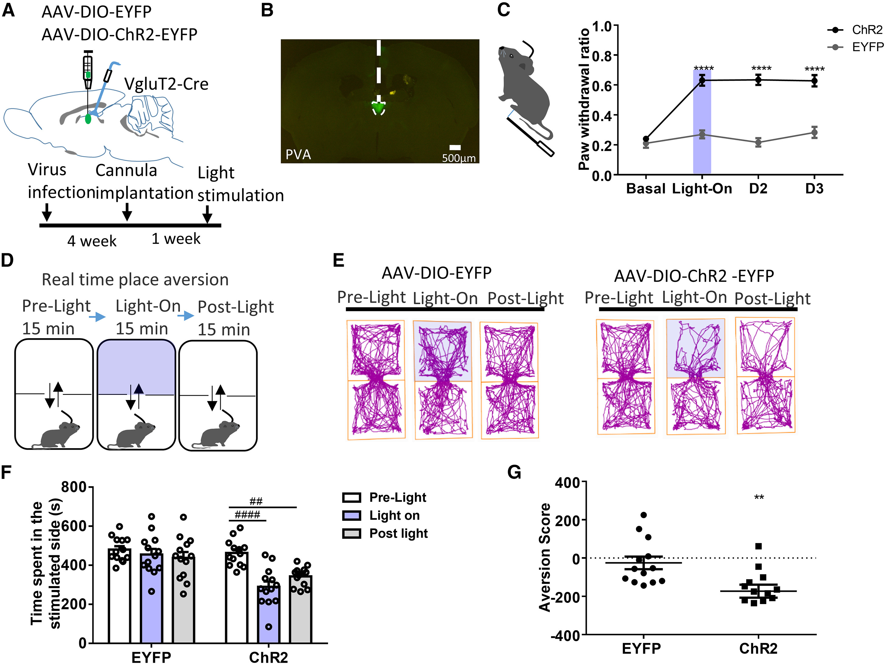
To test whether activating PVA VgluT2+ neurons is sufficient to drive nociceptive and affective pain behaviors, the authors expressed ChR2-EYFP in the PVA of naive VgluT2-ires-Cre mice. Strikingly, a single 10-minute bout of blue-light photostimulation (473 nm, 10 Hz) induced robust mechanical hypersensitivity (elevated PWR) that persisted for 3 days before returning to baseline by Day 6 (n = 15–16).
In the real-time place aversion (RTPA) assay, mice expressing ChR2 in PVA VgluT2+ neurons spent significantly less time in the photostimulation-paired chamber (n = 13, p < 0.001; chemogenetic hM3Dq activation similarly induced conditioned place aversion [CPA] in Figure S2A–E), demonstrating that PVA VgluT2+ activation suffices to trigger both persistent mechanical hypersensitivity and aversive affect.
Source: Selomon Assefa Mindaye et al. · Cell Reports · © Elsevier Inc.
Original figure and English caption ↗
Optogenetic Inhibition of PVA VgluT2+ Neurons Attenuates Formalin-Induced Mechanical Hypersensitivity and Drives Place Preference
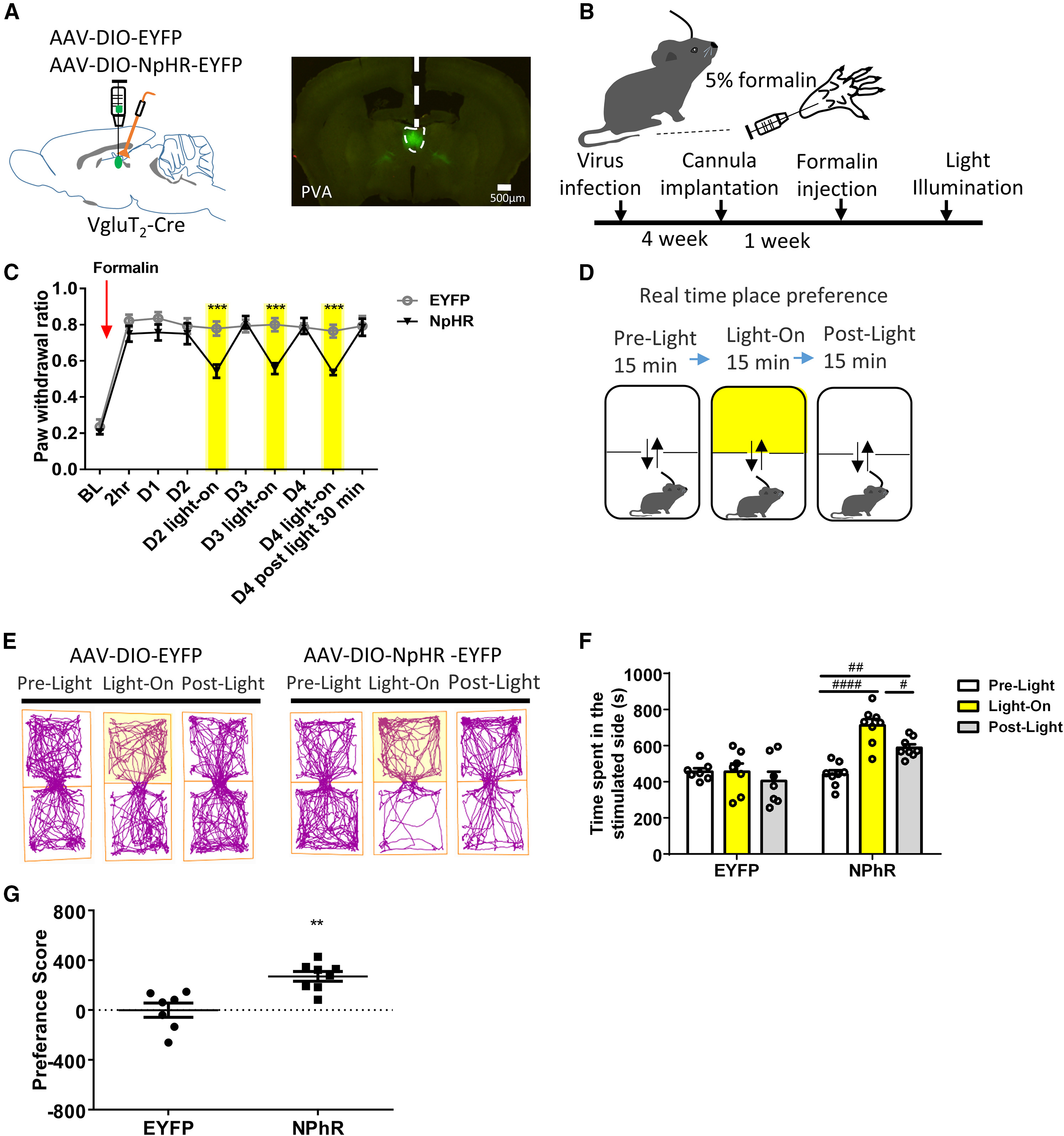
To test whether ongoing PVA VgluT2+ neuronal activity is required to maintain inflammatory pain behaviors, the authors expressed the inhibitory opsin eNpHR3.0-EYFP in the PVA of VgluT2-ires-Cre mice. Across 3 consecutive days following intraplantar formalin injection, yellow-light illumination (595 nm) reversibly suppressed mechanical PWR during light-on epochs, with hypersensitivity rebounding 1 hour after light termination (n = 7).
In affective assays, optogenetic inhibition of PVA VgluT2+ neurons in formalin-treated mice induced significant real-time place preference (RTPP) for the light-paired chamber (n = 7–8) and abolished formalin-induced conditioned place aversion (CPA; Figure S3A–D), confirming that PVA VgluT2+ activity is required for both sensory and affective pain dimensions.
Source: Selomon Assefa Mindaye et al. · Cell Reports · © Elsevier Inc.
Original figure and English caption ↗
Longitudinal Miniscope Imaging Reveals Non-overlapping PVA VgluT2+ Ensembles Encoding Mechanical Pain Versus Conditioned Aversion
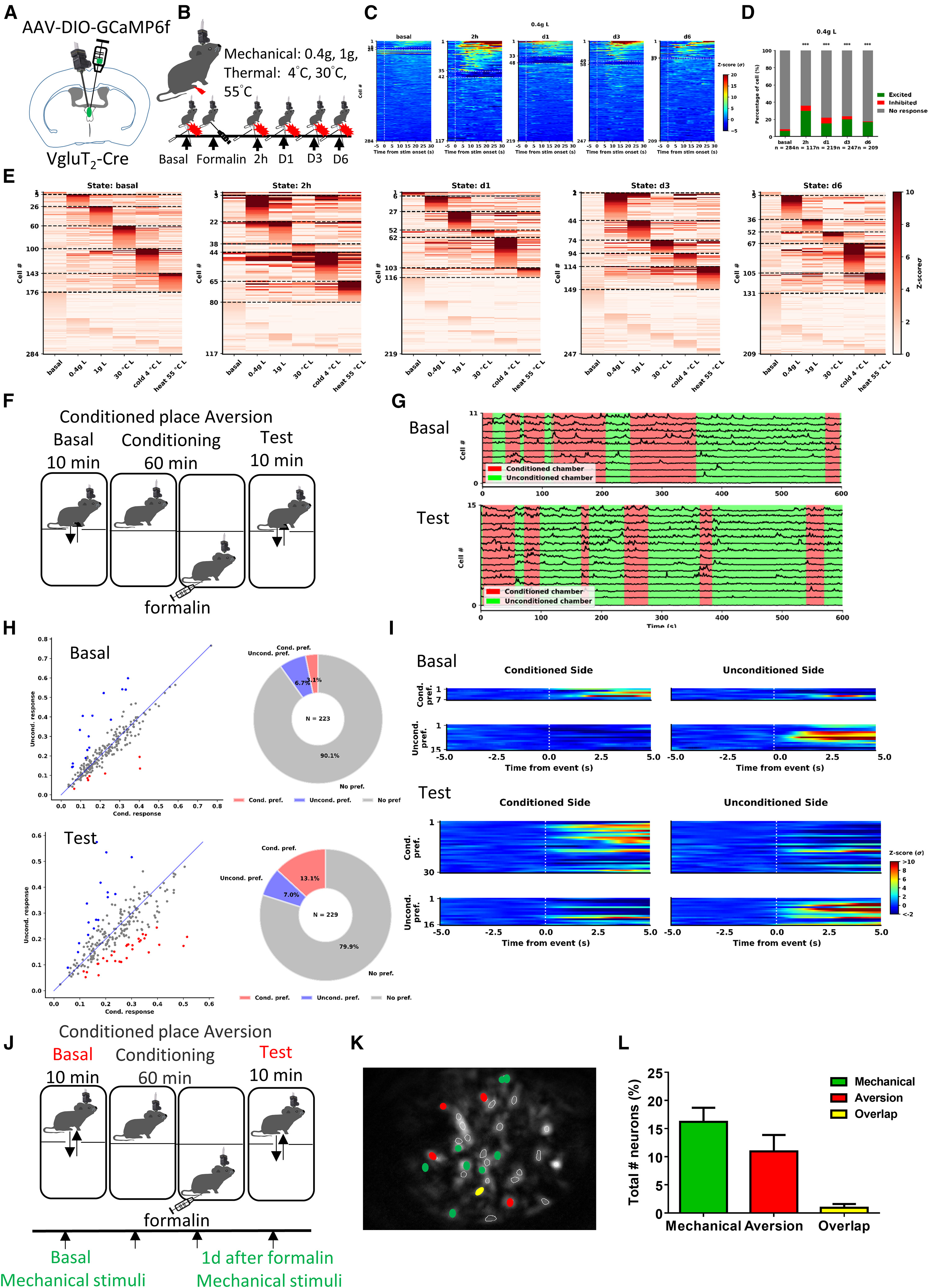
To determine whether individual PVA VgluT2+ neurons co-encode mechanical hypersensitivity and affective aversion, the authors performed longitudinal miniscope Ca2+ imaging across baseline and 2 hours, 1 day, 3 days, and 6 days post-formalin (n = 8 mice). Mechanical stimulation with a 0.4 g von Frey filament evoked significantly heightened Ca2+ transients in PVA VgluT2+ neurons at 2 hours, 1 day, and 3 days post-formalin, whereas responses to thermal stimuli (30 °C, 55 °C, and 4 °C) remained unchanged.
During formalin-induced conditioned place aversion (CPA), the proportion of PVA VgluT2+ neurons selectively responsive to the formalin-paired chamber (Cond. Pref) increased from 3.1% at baseline to 13.1% at test (unconditioned chamber: 6.7% vs 7.0%). Strikingly, cross-registration of 237 neurons across 8 mice revealed that CPA-responsive neurons (28 neurons, 12%) and mechanical-pain-responsive neurons (24 neurons, 10%) were virtually non-overlapping, with only 2 neurons (1%) responding to both (and anatomical tracing in Figure 5 identified the NAc and BNST as primary targets of nociception-activated PVA VgluT2+ neurons).
Source: Selomon Assefa Mindaye et al. · Cell Reports · © Elsevier Inc.
Original figure and English caption ↗
Optogenetic Activation of PVA VgluT2+→BNST Terminals Induces Persistent BNST Hyperactivity and Selectively Drives Mechanical Hypersensitivity
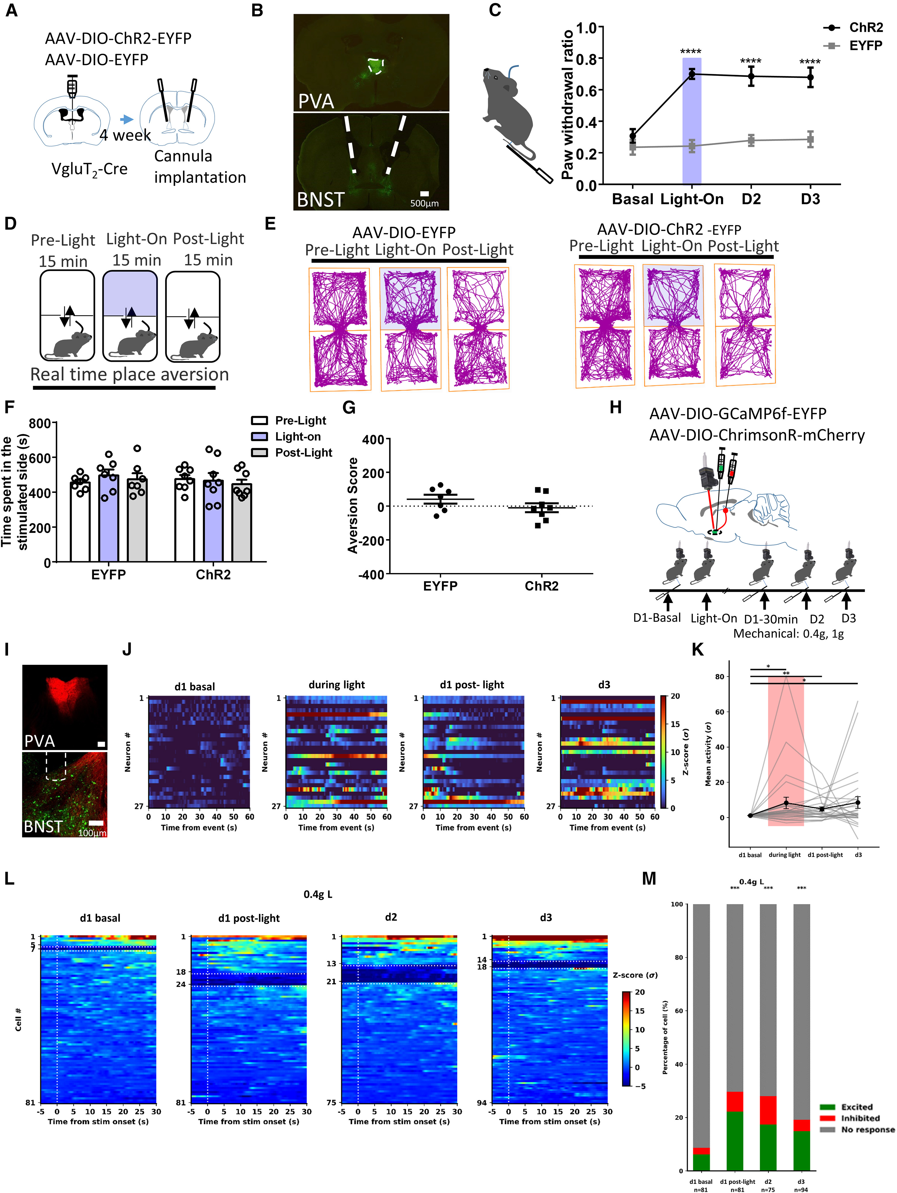
Guided by viral tracing showing that formalin-activated PVA VgluT2+ neurons project prominently to the bed nucleus of the stria terminalis (BNST) and nucleus accumbens (NAc) (Figure 5), the authors first optically stimulated ChR2-expressing PVA VgluT2+ axon terminals in the BNST. Terminal photostimulation in naive mice induced mechanical hypersensitivity (elevated PWR) lasting for at least 3 days (n = 7) without eliciting place avoidance in the RTPA test (n = 7–8).
To determine how brief terminal activation produces multi-day mechanical hypersensitivity, the team combined red-shifted ChrimsonR expression in PVA VgluT2+ neurons with GCaMP6f miniscope imaging in the BNST. Simultaneous 620 nm photostimulation of PVA VgluT2+→BNST terminals and BNST Ca2+ imaging revealed persistent increases in both spontaneous and 0.4 g von Frey-evoked Ca2+ activity in BNST neurons lasting up to 3 days post-stimulation.
Source: Selomon Assefa Mindaye et al. · Cell Reports · © Elsevier Inc.
Original figure and English caption ↗
The PVA VgluT2+→NAc Pathway Selectively Drives Place Avoidance and Is Recruited During Formalin CPA
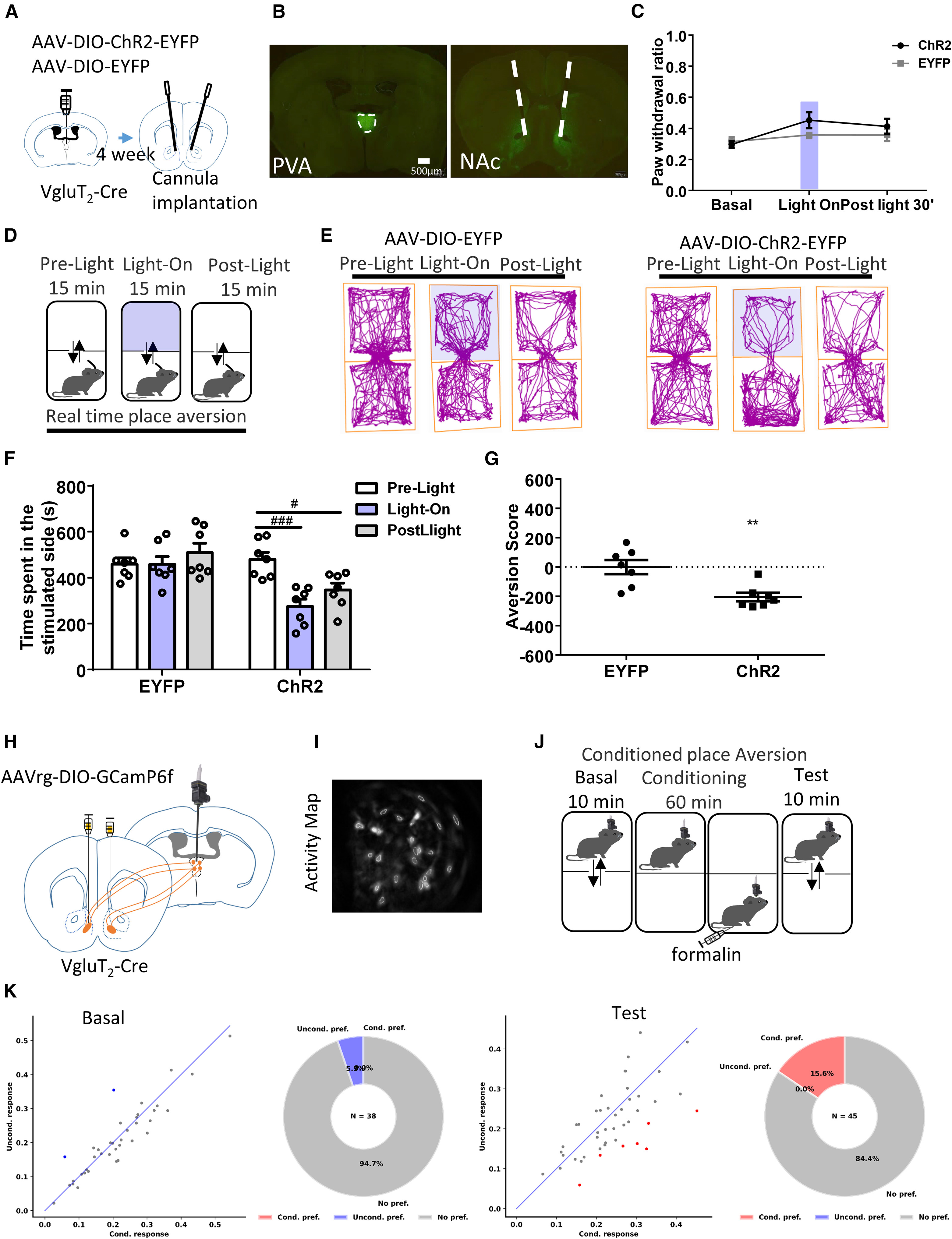
In mirror-image contrast to the BNST pathway, optogenetic stimulation of ChR2-expressing PVA VgluT2+ axon terminals in the nucleus accumbens (NAc) induced robust avoidance in the RTPA assay (n = 7, p < 0.01) while leaving mechanical PWR completely unaltered from 2 hours through 6 days post-stimulation (n = 7–8).
Finally, retrograde miniscope imaging of NAc-projecting PVA VgluT2+ neurons (via AAVrg-EF1α-DIO-GCaMP6f-tdTomato injected into the NAc and a GRIN lens in the PVA) demonstrated that the proportion of conditioned-chamber-responsive PVA VgluT2+→NAc neurons rose from 0% at baseline to 15.6% during the formalin CPA test. Together, these findings establish that segregated PVA VgluT2+→BNST and PVA VgluT2+→NAc projections independently govern the sensory-discriminative and affective-motivational dimensions of inflammatory pain.
Source: Selomon Assefa Mindaye et al. · Cell Reports · © Elsevier Inc.
Original figure and English caption ↗