How a three-stage motor-related network shapes pain-like responses
Nigral inhibitory input, subthalamic nucleus and lateral parabrachial nucleus form a modulatory route. Reducing downstream output or enhancing upstream input improves selected hypersensitivity measures in mouse inflammation and nerve-injury models.
Subthalamic nucleus (STN) is often associated with motor control, yet pain-related responses also alter its activity. How does this basal-ganglia region connect to bodily sensory signals? A study published in Nature Communications on December 15, 2022 follows a three-stage route from substantia nigra pars reticulata (SNr) through STN to lateral parabrachial nucleus (LPB). Upstream inhibitory input modulates STN, whose excitatory output reaches a pain-processing region. Inflammation and nerve injury shift inhibition and excitation in different directions, and counteracting these changes alters selected pain-like responses in mice.
The route modulates pain-like hypersensitivity. Removing LPB-projecting STN cells abolishes selected upstream-stimulation benefits while model-associated hypersensitivity can still develop.
Contents · 7 sections
STN activity changes during pain-like states
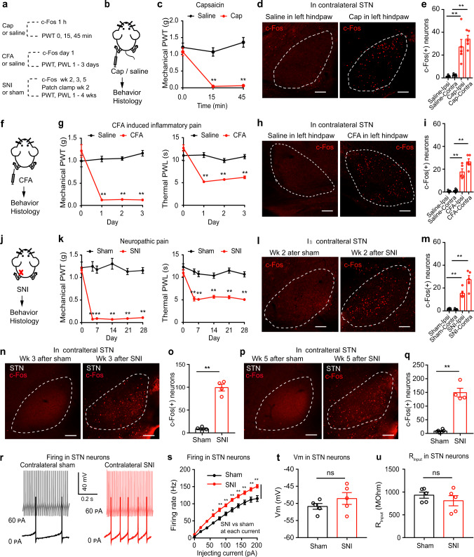
STN c-Fos-positive cells increase after acute capsaicin stimulation and in inflammatory and nerve-injury models, persisting weeks after injury. Slice STN cells fire more under the same current drive after nerve injury, without significant changes in resting potential or input resistance.
STN links a motor-related network with sensory regulation. Greater evoked firing can coexist with stable basic electrical properties, directing attention to the inputs these cells receive and the destinations of their output.
Source: Jia T et al. · Nature communications · CC BY
Original figure and English caption ↗
Excitatory output reaches a pain-processing brainstem node
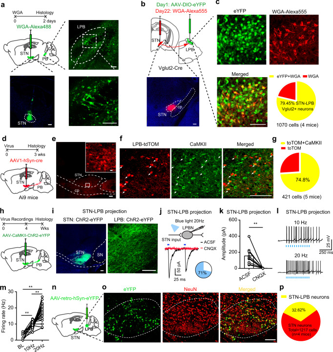
Tracing identifies STN-related input in LPB, with most recipient-labeled cells carrying glutamatergic-related markers. Activating STN terminals in slices evokes excitatory currents in a subset of LPB cells, reduced by glutamate-receptor blockade, and increases firing in responsive cells.
The connection includes both anatomical evidence and functional excitation. Recipients are predominantly glutamatergic-related but include other labeled types. This output provides a specific link through which basal ganglia can influence sensory responses.
Source: Jia T et al. · Nature communications · CC BY
Original figure and English caption ↗
Projection-defined cells affect mechanical and thermal measures differently
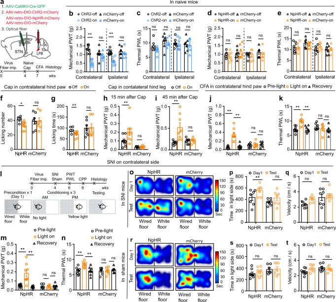
Activating LPB-projecting STN cells lowers bilateral mechanical withdrawal thresholds in naive mice without significantly changing thermal withdrawal latency. Inhibition leaves normal thresholds largely unchanged, but chiefly improves contralateral hypersensitivity in inflammation and nerve-injury models. Modality and laterality both matter.
Nerve-injured mice prefer the inhibition-paired environment, unlike sham controls, with no significant speed change there. The manipulation acts on projection-defined somata that have other outputs, motivating local-terminal tests in LPB.
Source: Jia T et al. · Nature communications · CC BY
Original figure and English caption ↗
Local terminals reveal a different scope
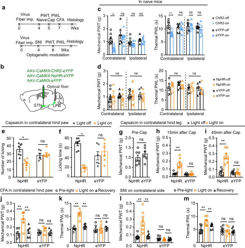
Activating STN terminals in LPB lowers contralateral mechanical thresholds and thermal latency in naive mice, without significant ipsilateral changes. Local inhibition again leaves normal thresholds largely unchanged. This differs from bilateral mechanical effects of soma activation, showing that a population and a local output segment are not interchangeable.
Local inhibition reduces contralateral nocifensive behavior or improves hypersensitivity thresholds in capsaicin, inflammatory and nerve-injury models. Related female-mouse model experiments show corresponding pathway effects. Responses to the same inhibition differ between naive and hypersensitive states.
Source: Jia T et al. · Nature communications · CC BY
Original figure and English caption ↗
Nerve injury shifts upstream inhibition and downstream excitation
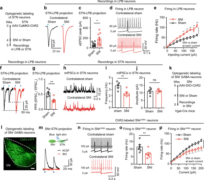
After nerve injury, STN-terminal-evoked LPB currents are larger and paired-pulse ratio is lower, supporting a presynaptic contribution to enhanced output. LPB evoked firing also increases. Meanwhile, STN miniature inhibitory-current frequency falls without a significant amplitude change.
Among candidate inputs, SNr GABAergic spontaneous and evoked firing decrease, unlike two other tested input regions. Less inhibition-related activity alongside stronger excitation supports a model of weakened braking and increased downstream responses.
Source: Jia T et al. · Nature communications · CC BY
Original figure and English caption ↗
Enhancing upstream input reduces LPB responses
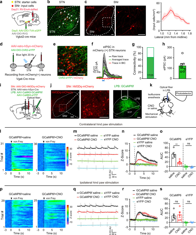
Tracing and slice recordings link inhibitory SNr input to LPB-projecting STN cells. Activating labeled SNr terminals evokes GABA-receptor-sensitive inhibitory currents in a subset of projection-defined cells, linking both segments through a relay population.
Increasing activity of STN-projecting SNr cells reduces LPB calcium responses to contralateral mechanical stimulation, without a significant ipsilateral change. The recording parallels the laterality of earlier effects and supports upstream modulation of downstream sensory responses.
Source: Jia T et al. · Nature communications · CC BY
Original figure and English caption ↗
Removing the relay abolishes benefit while hypersensitivity still develops
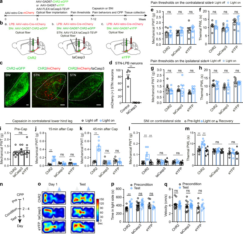
Activating inhibitory SNr terminals in STN improves selected contralateral thresholds in capsaicin and nerve-injury models and produces paired-environment preference in injured mice. Removing LPB-projecting STN cells abolishes these upstream-stimulation benefits and preference effects, with largely unchanged baseline thresholds and motor comparisons.
Relay-ablated mice still develop model-associated hypersensitivity. This separates the relay supporting upstream-stimulation benefits from initiation of hypersensitivity. Tested adult male and female results reveal state-dependent basal-ganglia modulation of sensory responses within a wider network.
Source: Jia T et al. · Nature communications · CC BY
Original figure and English caption ↗