From deep insula to thalamus: a connection shaping sensitivity and avoidance
A mouse study connects cortical layers, thalamic location and downstream cells, finding different effects of the same pathway at baseline and after nerve injury.
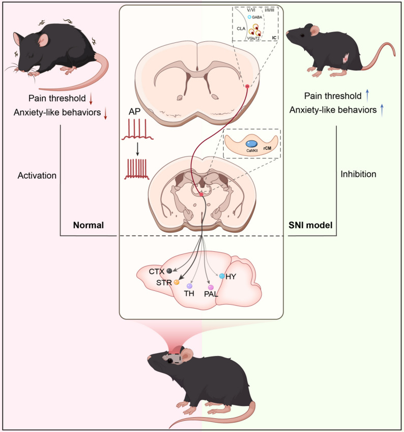
Pain can change responses to stimulation and willingness to explore open spaces. How are these changes connected in the brain? A 2026 The Journal of Headache and Pain study in the spared nerve injury, or SNI, mouse model focuses on an excitatory connection between deep insular layers five and six, or dlIC, and rostral central medial thalamus, or rCM. Moving from regions into projecting cells, terminals and input recipients, the study finds different regulatory effects at baseline and after injury.
dlIC glutamatergic cells and their rCM connection contribute to sensitivity and avoidance. Activation produces related changes in controls, suppression improves SNI outcomes, and baseline suppression is largely without significant effects.
Contents · 6 sections
Functional differences exist within insula and thalamus
Post-injury activity labeling emphasized deep insular layers and rostral CM, mainly with excitatory markers. Comparing cortical layers, rostrocaudal thalamic positions and adjacent nuclei narrowed two broad regions into a more defined connection. Pain-associated recruitment had a particular distribution within each region.
Retrograde labeling identified dlIC input to rCM, and anterograde terminals approached CaMKIIα-associated rCM cells. Following this direction, the researchers examined projecting-cell physiology and defined downstream input recipients. The anatomical map thus led into activity and behavior.
Projecting cells become more readily recruited
After SNI, rCM-projecting dlIC cells received more frequent and larger excitatory inputs, had depolarized resting potentials and lower rheobase, and fired more. Some spike parameters remained similar, while paired-pulse changes supported release-related effects. Inputs and cellular responsiveness thus changed together.
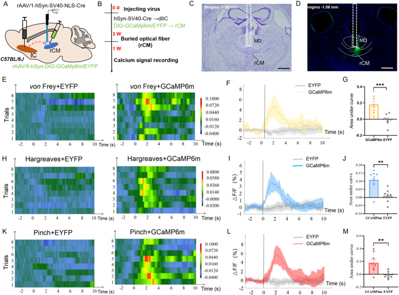
Mechanical, thermal and tail-pinch stimuli also recruited dlIC-recipient rCM cells in naïve mice. The connection was active during noxious stimulation, and projecting cells were more excitable after injury. These observations led to tests of how modulation affects thresholds and exploration.
Source: Ren X et al. · The journal of headache and pain · CC BY-NC-ND
Original figure and English caption ↗
Activation in controls, suppression after injury
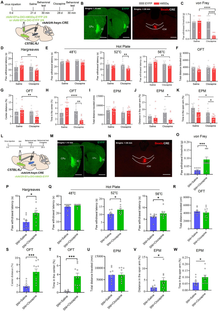
Increasing rCM-projecting dlIC activity in controls lowered mechanical and thermal thresholds and reduced center and open-arm exploration, with similar overall locomotion. Suppression at baseline produced largely nonsignificant sensory and behavioral changes. The differing effects clarified the connection’s state dependence.
Similar total locomotor distance with less center and open-arm exploration describes a change in how animals allocate movement across space. Lower mechanical and thermal thresholds show altered responses to two stimulus types. Examining these measures together captures both sensory responses and choices of where to move.
Suppressing related activity in SNI mice raised thresholds and improved avoidance. Smaller baseline effects and clearer post-injury effects aligned with injury-associated cellular recruitment. The connection’s influence depended on network state, with different regulatory relationships at baseline and after injury.
Source: Ren X et al. · The journal of headache and pain · CC BY-NC-ND
Original figure and English caption ↗
Terminals and recipient cells show convergent effects
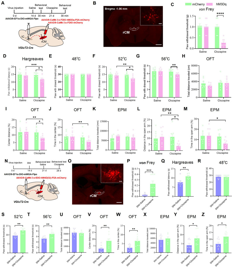
Modulating dlIC VGLUT2 terminals in rCM produced sensitivity enhancement in controls or improvement after injury. Activation of defined CaMKIIα-associated recipients lowered thresholds and exploration, while suppression improved SNI outcomes. Findings converged from upstream cells through terminals to the downstream population.
Following the connection made the functional relationship between the regions more specific. Convergent effects in a population receiving defined input located sensory and behavioral changes within a traceable network segment.
Source: Ren X et al. · The journal of headache and pain · CC BY-NC-ND
Original figure and English caption ↗
Input recipients have different output patterns
Three-dimensional reconstructions identified restricted and broader output patterns in dlIC-recipient rCM-related cells, with the latter reaching cortical and striatal regions. After thalamic reception, signals could enter different networks. These morphological differences added spatial information about subsequent propagation.
Functional findings concern the recipient population, while the morphological classes have not been separately assigned behavioral roles. Relating their outputs to thresholds and exploration will be key to understanding effects across readouts. A shared input source with different destinations makes organization within the thalamus an important question.
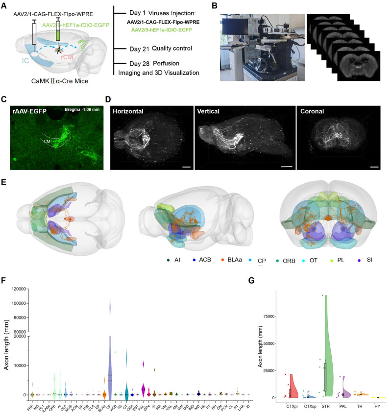
Source: Ren X et al. · The journal of headache and pain · CC BY-NC-ND
Original figure and English caption ↗
Joint threshold and exploration changes identify a regulatory site
Withdrawal and thermal responses describe noxious-stimulus-related behavior, while open-space exploration also involves risk and action strategy. Joint changes under modulation of one connection identify dlIC→rCM as a site linking sensation with behavior. How pain relief influences exploration, or how different outputs contribute to each outcome, can be distinguished within this defined connection.
The study used male mice in the SNI model. Its localization shows the value of examining positions and input-recipient populations within a region separately. Narrowing a pain-associated state from regions into a connection, then placing it within a network of diverse outputs, is the study’s specific perspective.