After nerve injury, two thalamic regions diverge
A male mouse study separates persistent mechanical sensitivity from affect-related behavior: posterior paraventricular-thalamic activity rises while anterior activity falls, with different output pathways contributing.
After nerve injury, mechanical sensitivity can increase alongside changes in exploration and reward preference. Do these accompanying changes arise from the same neurons? A study published in Advanced Science on July 7, 2024 follows the paraventricular thalamus in male mice and finds opposing activity changes at its anterior and posterior ends. Tracking their outputs distinguishes pathways associated with mechanical sensitivity and affect-related behavior.
Suppressing posterior paraventricular thalamus and its ventrolateral periaqueductal-gray pathway improves post-injury mechanical thresholds. Increasing anterior-thalamic or related accumbal activity improves several affect-related behavioral measures.
Contents · 8 sections
Opposing changes within one brain region
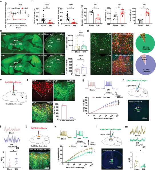
Hindpaw mechanical thresholds decrease after nerve injury and remain low through observation. By week six, mice spend less time in exposed spaces, prefer sucrose less, and show longer immobility in tail-suspension and swim tests. The study examines sensory responses alongside several affect-related measures.
Posterior paraventricular-thalamic, or PVP, excitatory cells spike more readily and show greater population calcium activity, whereas anterior PVA firing and calcium activity decline. Neighboring regions change in opposite directions, suggesting different behavioral contributions.
Source: Deng J et al. · Advanced science (Weinheim, Baden-Wurttemberg, Germany) · CC BY
Original figure and English caption ↗
Reducing posterior activity raises mechanical thresholds
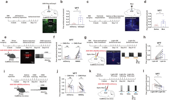
Reducing PVP excitatory-cell activity raises mechanical thresholds in injured mice. Conversely, activating these cells in uninjured mice lowers thresholds. Different interventions converge on a contribution of higher PVP activity to mechanical sensitivity.
Reduced mechanical sensitivity does not accompany uniform improvement across affect-related measures. Sucrose preference and tested suspension/swim measures remain broadly unchanged after PVP suppression, while one center-exploration measure increases after cell ablation. The sensory and behavioral effects show distinguishable emphases.
Source: Deng J et al. · Advanced science (Weinheim, Baden-Wurttemberg, Germany) · CC BY
Original figure and English caption ↗
Recruiting anterior cells changes exploration and reward preference
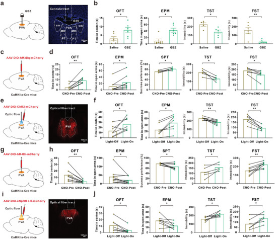
PVA shows the opposite pattern. Increasing its excitatory-cell activity increases center and open-arm time, raises sucrose preference and reduces immobility in injured mice. Mechanical thresholds do not significantly change despite these behavioral effects.
Suppressing PVA in uninjured mice reduces exposed-space exploration and changes some reward/immobility measures without the corresponding mechanical sensitization. Opposing interventions support different anterior and posterior contributions, allowing distinct post-injury outcomes to be regulated separately.
Source: Deng J et al. · Advanced science (Weinheim, Baden-Wurttemberg, Germany) · CC BY
Original figure and English caption ↗
Inhibitory cells can contribute to increased sensitivity
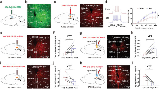
PVP outputs reach ventrolateral periaqueductal gray, or VLPAG, in the midbrain. After injury, its GABA cells spike more readily. Inhibiting them raises mechanical thresholds; activating them in uninjured mice lowers thresholds.
Using an inhibitory transmitter does not mean that increasing a cell’s activity necessarily reduces sensitivity. The targets inhibited and the cell’s network position shape the behavioral outcome. In this model, more active VLPAG GABA cells are associated with lower mechanical thresholds.
Source: Deng J et al. · Advanced science (Weinheim, Baden-Wurttemberg, Germany) · CC BY
Original figure and English caption ↗
A thalamic–midbrain connection changes behavior
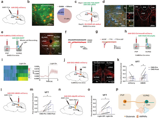
Tracing and slice recordings connect these populations. PVP terminals evoke excitatory currents in VLPAG GABA cells, with pharmacological tests supporting direct synaptic input. An excitatory output can therefore recruit downstream inhibitory cells.
Reducing PVP terminal input or suppressing VLPAG cells receiving it raises mechanical thresholds in injured mice. These recipients include many GABA cells and other cell types. The downstream population identified through thalamic input thus contributes to regulation of mechanical sensitivity.
Source: Deng J et al. · Advanced science (Weinheim, Baden-Wurttemberg, Germany) · CC BY
Original figure and English caption ↗
Anterior outputs reach accumbal cells
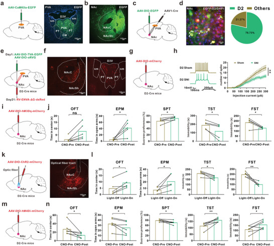
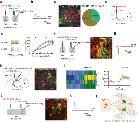
Following PVA outputs leads to nucleus accumbens. Its cells are often distinguished by D1 or D2 dopamine-receptor expression, and D2 cells receive PVA input. Injured mice show weaker D2 firing and calcium activity, matching the direction of lower PVA activity.
Activating D2 cells improves sucrose preference and immobility measures, while center-exploration findings differ across activation experiments. Sustained D2 inhibition produces opposing changes in several measures in uninjured mice. Receptor names identify the cell populations here; the interventions alter cellular activity.
Source: Deng J et al. · Advanced science (Weinheim, Baden-Wurttemberg, Germany) · CC BY
Original figure and English caption ↗
Recipient populations influence affect-related behavior
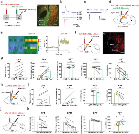
Recruiting PVA terminals evokes excitatory currents in D2 cells, with tests supporting direct input, and increases D2 population calcium activity. Anterior-thalamic output can transmit signals to accumbens rather than simply changing in parallel with it.
Activating accumbal cells receiving PVA input improves exploration, sucrose preference and immobility measures in injured mice; inhibiting this population alters behavior in uninjured mice. The recipient population includes multiple cell types, prompting examination of how they communicate locally.
Source: Deng J et al. · Advanced science (Weinheim, Baden-Wurttemberg, Germany) · CC BY
Original figure and English caption ↗
Alongside direct excitation, a local inhibitory branch
D2 cells also receive inhibitory currents. Further recordings show that PVA excites D1 cells, which can provide GABAergic inhibition to D2 cells. Thalamic input thus enters an accumbal network containing direct excitation and a local inhibitory branch.
Separately manipulating PVA-recipient D1 cells does not significantly change the tested behaviors. This adult male mouse study reveals circuit complexity while relating persistent mechanical sensitivity and affect-related responses to different output pathways. Co-occurring outcomes can have distinct regulatory components.
Source: Deng J et al. · Advanced science (Weinheim, Baden-Wurttemberg, Germany) · CC BY
Original figure and English caption ↗