Why electrical stimulation and cell activation affect seizures differently
A male-mouse motor-cortex seizure model shows improved EEG and seizure measures with tested high-frequency subthalamic stimulation. Direct excitatory-cell activation worsens seizures; nigral input and orexin receptors provide mechanistic clues.
At one brain site, electrical stimulation improves seizures while directly activating excitatory cells worsens them. Understanding stimulation requires examining its network effects. A study published in Frontiers in Neuroscience on May 5, 2023 tests subthalamic nucleus, STN, and its output to substantia nigra pars reticulata, SNr, in a male-mouse motor-cortex seizure model. Within the basal-ganglia network, STN excitatory outputs reach SNr, a region dominated by GABAergic cells. Signals passing through different cells and destinations can influence seizure propagation, making output destination important. Population, terminal and orexin-receptor experiments investigate circuit and molecular clues to the improvement.
Tested high-frequency STN stimulation reduces EEG spikes and selected seizure measures. Inhibiting STN excitatory cells or their SNr terminals also improves seizures. Activating SNr-projecting cells weakens stimulation benefits, while dual nigral orexin-receptor blockade improves several seizure measures.
Contents · 7 sections
Different seizure measures respond differently
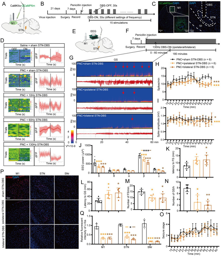
After motor-cortex seizure induction, the tested higher-frequency STN stimulation reduces cortical calcium responses, unlike lower and intermediate frequencies. Ipsilateral and bilateral high-frequency stimulation reduce EEG spike frequency, amplitude and selected spectral power, along with focal and generalized seizure counts.
Latency measures differ: ipsilateral stimulation delays generalized seizures, while bilateral stimulation also delays focal onset. Neither prevents behavioral-stage progression in this comparison. Onset, number and progression therefore represent distinct effects.
Source: Xue T et al. · Frontiers in neuroscience · CC BY
Original figure and English caption ↗
Direct excitatory-cell activation intensifies seizures
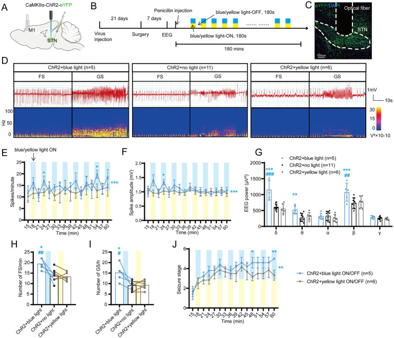
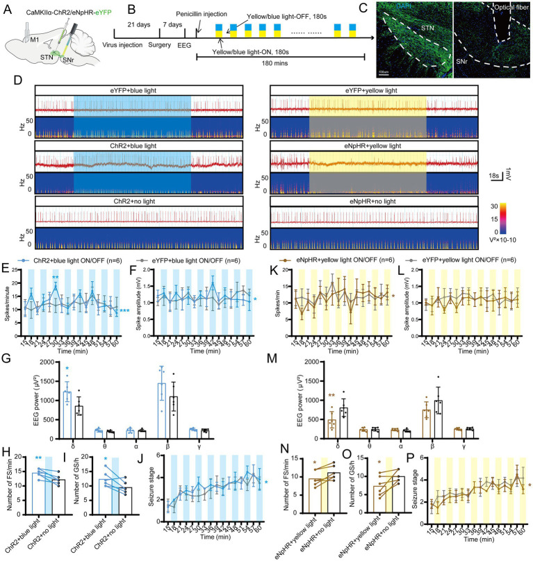
Most STN cells are glutamatergic. Direct optical activation of the labeled excitatory population increases EEG spike frequency and amplitude, focal and generalized seizure counts, and the rate of behavioral-stage progression. Control conditions lack the same changes.
Electrical stimulation and cellular activation act in different directions. High-frequency stimulation may alter inputs, outputs and nearby fibers, motivating circuit-level interpretation. Increasing this excitatory population promotes seizures in the model.
Source: Xue T et al. · Frontiers in neuroscience · CC BY
Original figure and English caption ↗
Inhibiting the population shifts effects in the opposite direction
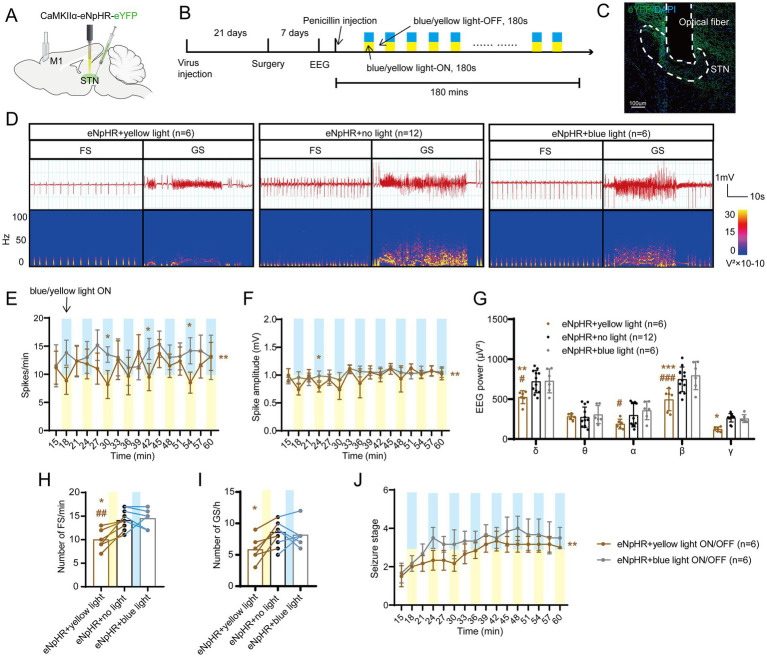
Reducing STN excitatory activity lowers EEG spike frequency and amplitude, selected spectral power and focal seizure count. Generalized seizures also decrease versus no-light conditions. Behavioral progression slows, supporting bidirectional population effects on seizure intensity.
This direction resembles some high-frequency stimulation effects and motivates tracing STN output. SNr receives STN input and is a basal-ganglia node involved in seizure regulation. The study next focuses on this connection.
Source: Xue T et al. · Frontiers in neuroscience · CC BY
Original figure and English caption ↗
Projection-defined cells also influence seizures
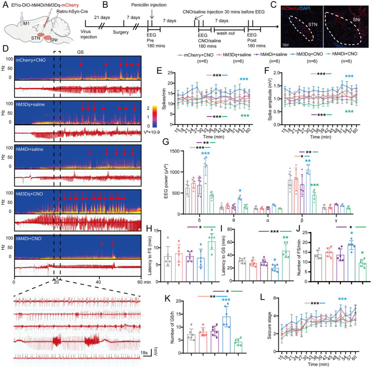
Inhibiting STN cells defined by projection to SNr reduces spikes and seizure counts and delays focal and generalized onset, without significantly slowing behavioral-stage progression. Activating the population increases abnormal discharges and seizures and accelerates progression.
Activation more clearly shortens generalized-seizure latency; the main focal-onset comparison is not significantly shorter. The manipulation acts on STN somata that may have other branches, motivating local-terminal tests in SNr.
Source: Xue T et al. · Frontiers in neuroscience · CC BY
Original figure and English caption ↗
Local terminals identify an output contribution
Activating STN terminals in SNr likewise increases spikes, seizure counts and progression. Terminal inhibition lowers spike frequency and selected spectral power, reduces seizures and slows progression, without a significant spike-amplitude reduction.
Population and terminal experiments support a shared overall direction while retaining differences among measured outcomes. They link STN-to-SNr output with seizure regulation and motivate testing its contribution to electrical-stimulation effects.
Source: Xue T et al. · Frontiers in neuroscience · CC BY
Original figure and English caption ↗
Increasing output weakens stimulation-related improvement
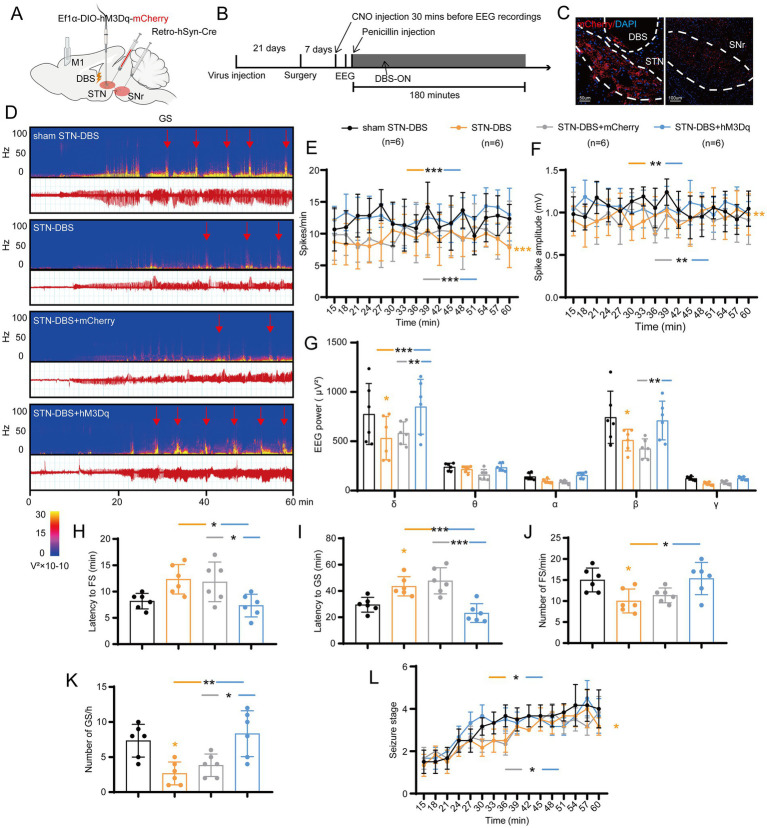
During high-frequency STN stimulation, activating SNr-projecting STN cells raises spike frequency, amplitude and spectral power relative to the improved conditions and weakens several generalized-seizure benefits. Marker controls retain the stimulation-associated direction of improvement.
The combined experiment supports involvement of STN-to-SNr activity changes in stimulation effects. It does not directly track every terminal’s firing during stimulation, and other STN outputs may contribute. The findings identify a more specific circuit component.
Source: Xue T et al. · Frontiers in neuroscience · CC BY
Original figure and English caption ↗
Orexin receptors separate EEG and behavioral effects
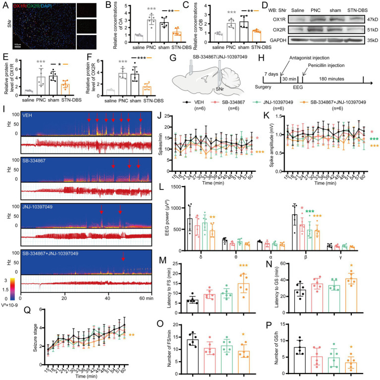
SNr tissue orexin A and B and both receptor proteins rise in the seizure model and decline after stimulation. OX1R blockade improves spike frequency, amplitude and selected power. OX2R blockade improves amplitude and power without significantly lowering spike frequency.
Clear behavioral benefits chiefly appear with dual blockade: onset is delayed, counts fall and progression slows, unlike nonsignificant single-blockade comparisons. These acute male-mouse motor-cortex findings link circuit regulation with local peptide-receptor action. Tissue changes accompany stimulation effects, while orexin’s necessity as their mediator is not separately tested.
Source: Xue T et al. · Frontiers in neuroscience · CC BY
Original figure and English caption ↗