How stress increases vulnerability in a motor-related system
Direct social stress worsens selected motor deficits and nigral TH-positive-cell reductions after MPTP in male mice. Amygdalar input and related downstream-cell activity contribute to vulnerability.

Can prior stress change how the brain responds to the same damaging exposure? A study published in CNS Neuroscience & Therapeutics on March 13, 2023 applies social stress before MPTP in male mice, then examines motor performance and nigral cell markers. MPTP models are widely used to investigate Parkinson-related motor-system injury. Direct social defeat increases sensitivity to selected toxin-associated outcomes. The team follows connections between the central amygdala (CeA) and substantia nigra pars compacta (SNc) to investigate where stress information meets motor-system vulnerability.
Stress experience, activity related to CeA input and manipulation of labeled SNc cells converge on vulnerability. Interventions occur during stress or toxin exposure.
Contents · 6 sections
Direct and witnessed stress produce different profiles
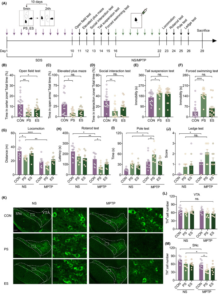
The team compares mice undergoing direct social attack, those witnessing another mouse’s attack and controls. Both stressed groups show altered open-field center time and social approach. Directly stressed mice also spend less time in elevated-plus-maze open arms and show more immobility in tail suspension and forced swimming.
Witnessed-stress mice do not differ significantly in the elevated-maze or immobility comparisons. Direct stress produces both avoidance and immobility changes, while witnessed-stress effects concentrate more on center time and social approach. The experiences leave different combinations of behavior. The distinction allows a question about which stress background accompanies greater subsequent motor impairment.
Source: Cai H et al. · CNS neuroscience & therapeutics · CC BY
Original figure and English caption ↗
Toxin exposure reveals differences in vulnerability
After MPTP, directly stressed mice fall sooner from the rotarod and perform worse in the pole test than controls receiving the same toxin. SNc TH-positive-cell counts are lower, while counts in the nearby ventral tegmental area (VTA) do not differ significantly. The rotarod examines balance and coordination, while open-field tracking describes spontaneous locomotion, offering different views of motor performance. TH is a dopamine-related marker, and the counts describe observed marker-positive cells.
Witnessed-stress mice do not show similarly clear toxin-associated differences in the tested motor measures and SNc counts. Motor outcomes are not uniform: the ledge test does not show further worsening by direct stress. Vulnerability appears in particular measures rather than every movement deteriorating together.
Following upstream input brings the amygdala into view
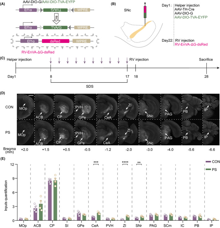
Tracing upstream from TH-related SNc cells reveals more labeled cells in CeA, zona incerta and substantia nigra pars reticulata after direct stress. Because CeA participates in stress and aversive processing, the team focuses on it. Increased labeling suggests a change in the mapped upstream-input pattern related to SNc.
The labeling map places stress-associated changes in upstream cells connected to SNc targets. Changes in zona incerta and nigral reticulata also indicate other network participants. The study then tests CeA-related connections without manipulating every altered input.
Source: Cai H et al. · CNS neuroscience & therapeutics · CC BY
Original figure and English caption ↗
Related signals increase during encounter with an aggressor
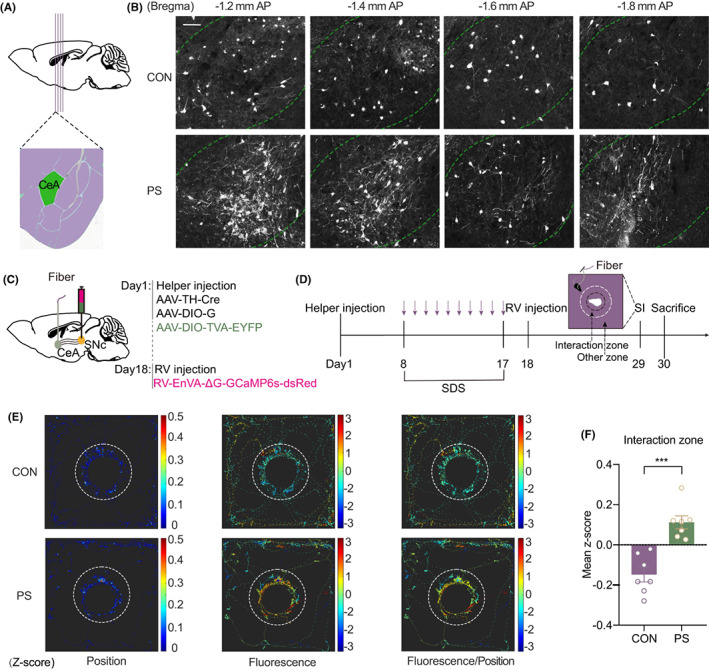
The team records CeA cells linked to TH-related SNc targets. During a social test with an aggressive mouse in the interaction area, directly stressed animals show higher average calcium signals corresponding to that area than controls. Activity in this input-related population thus changes when previously defeated animals encounter such a social target again.
The recordings bring social target, animal position and related cell activity into the same experience. The next experiments manipulate CeA-input-related labeled cells within SNc and examine motor performance and cell counts.
Source: Cai H et al. · CNS neuroscience & therapeutics · CC BY
Original figure and English caption ↗
Increasing downstream activity worsens selected outcomes
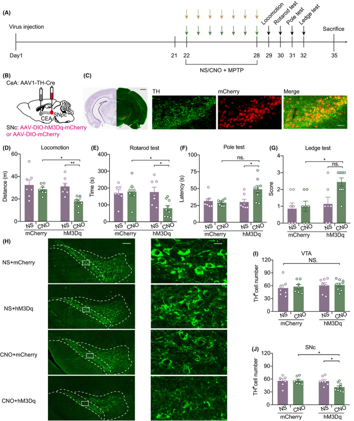
In mice without this social-stress regimen, the team increases activity of CeA-input-related labeled SNc cells during toxin exposure. Locomotor distance and rotarod latency decrease, SNc TH-positive counts are lower and VTA counts remain largely unchanged. This direction echoes selected outcomes after direct stress.
The manipulation acts on downstream SNc cells, whereas the recording experiment observes the upstream CeA population. Together they support involvement of this connection in vulnerability, without directly measuring pathway-wide synaptic currents or resolving each recipient cell type.
Source: Cai H et al. · CNS neuroscience & therapeutics · CC BY
Original figure and English caption ↗
Reducing activity during stress yields selective improvement
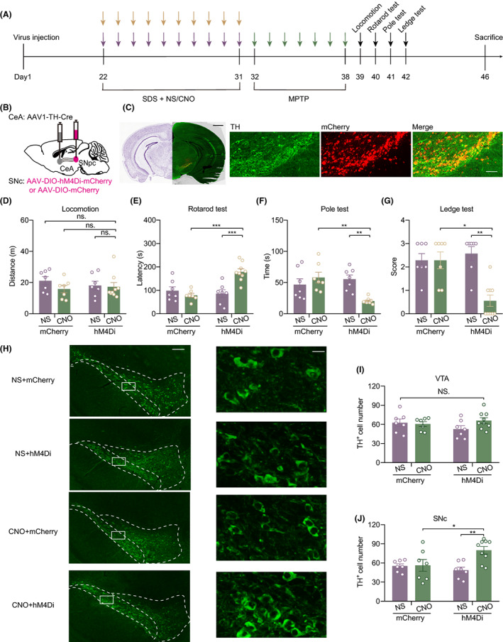
The reverse experiment inhibits the related labeled SNc population during social-stress development, before MPTP. Later, mice remain longer on the rotarod, improve in pole and ledge tests and retain more TH-positive SNc cells. VTA counts do not change significantly, nor does locomotor distance significantly recover.
Selective improvements distinguish coordination, locomotor distance and tissue markers, and place the intervention during stress development rather than after injury. The study uses adult male mice and excludes stress-resilient individuals in social testing. Its conclusions concern this stress-and-toxin model, offering experimental evidence on how emotion-related input influences motor-system vulnerability.
Source: Cai H et al. · CNS neuroscience & therapeutics · CC BY
Original figure and English caption ↗