Why do two thalamic branches shift seizures in opposite directions?
Calretinin-labeled neurons in one thalamic region have opposing circuit contributions through projections to the lateral amygdala and zona incerta. In mice, the consequences of targeting a region depend on its outputs.
Reducing activity in neurons recruited during seizures might seem certain to limit abnormal propagation. But what if the population sends outputs with opposing effects? A study published in iScience on April 7, 2022 examines this question in the posterior intralaminar thalamic nucleus (PIL). In a hippocampal kindling model in male mice, calretinin-labeled (CR) PIL neurons contribute to seizure development, while their branches to the lateral amygdala (LA) and zona incerta (ZI) have opposing circuit contributions.
Activating PIL CR neurons accelerates kindling, whereas inhibition slows it. Inhibiting the PIL-to-LA branch also slows development, but inhibiting the PIL-to-ZI branch accelerates it. Projection populations partially overlap, emphasizing the importance of downstream outputs.
Contents · 7 sections
A thalamic population becomes more active as seizures generalize
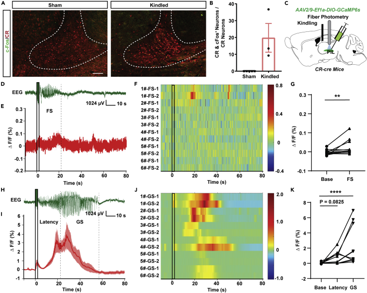
CR is a calcium-binding protein that labels different cellular populations across brain regions. In the kindling model, CR-cell numbers do not significantly change across surveyed regions, but the fraction of PIL CR neurons bearing the activity marker c-Fos increases. The detectable shift concerns activity rather than cell number.
Calcium recordings reveal a smaller increase during focal seizures and a much larger response during generalized seizures, with the peak near generalization onset. This identifies PIL as a candidate node and raises a causal question: is it merely recruited, or can it influence seizure development?
Source: Qi Y et al. · iScience · CC BY-NC-ND
Original figure and English caption ↗
Increasing activity speeds progression toward generalization
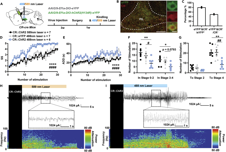
Activating PIL CR neurons accelerates progression toward higher seizure stages and reduces the number of stimulations required for the first generalized seizure. Electrographic afterdischarges also last longer, linking the behavioral progression to persistence of abnormal electrical activity.
Comparison groups vary illumination and include a fluorescent-marker-only condition to distinguish cellular activation from light exposure. The findings show that this PIL CR population can promote seizure development in the tested model rather than merely displaying activity after seizures.
Source: Qi Y et al. · iScience · CC BY-NC-ND
Original figure and English caption ↗
Suppressing the population mainly slows early development
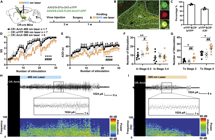
Suppressing the PIL CR population slows seizure development, shortens afterdischarges and increases the stimulation count required to reach seizure stages. Effects are more evident early; counts within higher stages show no equally clear change. The population-level result establishes a direction without identifying the relevant downstream route.
This motivates examining outputs separately. A population-level effect can combine multiple branches, and branches need not act in concert. Somatic activity alone may therefore obscure important circuit differences.
Source: Qi Y et al. · iScience · CC BY-NC-ND
Original figure and English caption ↗
One origin connects to several downstream regions
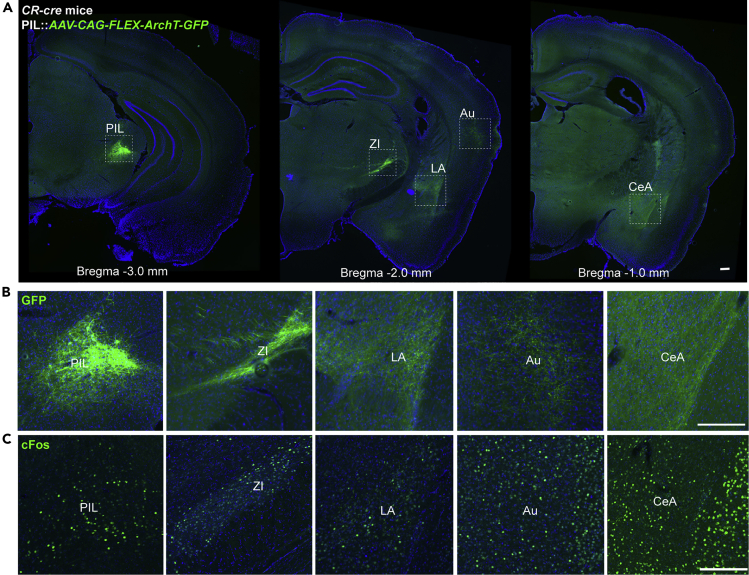
Most PIL CR neurons bear an excitatory-cell marker, with almost no colabeling for the tested GABAergic marker. Their axons reach LA, ZI, auditory cortex and central amygdala. LA and ZI attract particular attention because both receive prominent projections and show activity markers after generalized seizures.
These CR cells are not simply local inhibitory interneurons. They send information to distant targets, where the identity and connectivity of recipient cells can shape the eventual network consequence of an excitatory projection.
Source: Qi Y et al. · iScience · CC BY-NC-ND
Original figure and English caption ↗
Suppressing the amygdala branch slows development
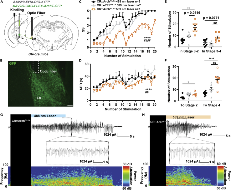
Inhibiting PIL CR terminals in LA slows seizure-stage progression, shortens afterdischarges and increases the number of stimulations needed for generalization. Examining this branch separately identifies an output contributing to seizure promotion.
In fully kindled mice, the effect has a different emphasis: generalized seizures are shorter and begin later, while seizure stage and total afterdischarge duration do not significantly change. The same projection therefore influences different measures during acquisition and after kindling is established.
Source: Qi Y et al. · iScience · CC BY-NC-ND
Original figure and English caption ↗
Suppressing the other branch has the opposite consequence
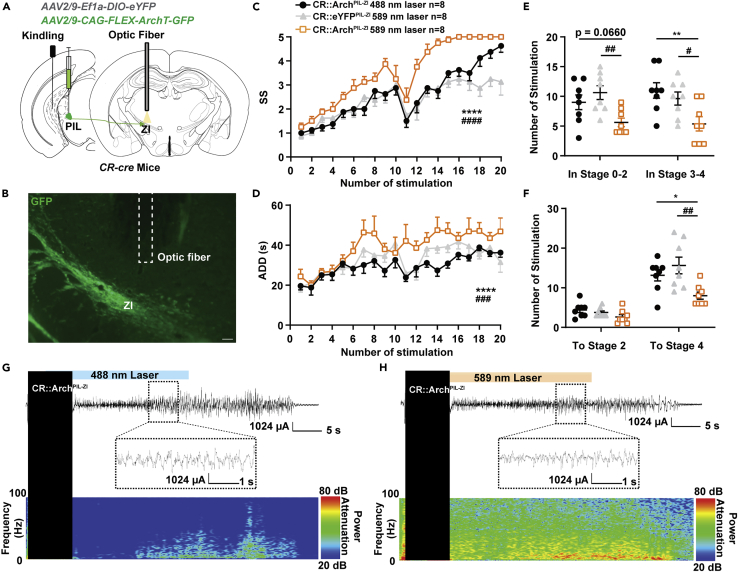
Applying the same strategy to ZI reverses the outcome. Inhibiting PIL-to-ZI terminals accelerates kindling, prolongs afterdischarges and brings the first generalized seizure earlier. This supports a restraining contribution of the branch within the tested network, so suppressing it removes part of that restraint.
After full kindling, generalized seizure duration increases, while seizure stage, total afterdischarge duration and latency to generalization remain unchanged. The branches differ both in their effects on development and on the duration of established generalized seizures.
Source: Qi Y et al. · iScience · CC BY-NC-ND
Original figure and English caption ↗
The two outputs share part of their source population
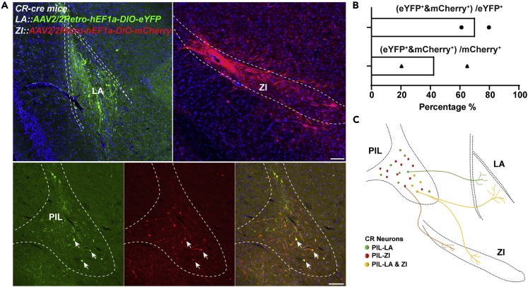
Dual retrograde labeling reveals partial overlap between LA-projecting and ZI-projecting CR neurons, suggesting that some cells connect to both through collaterals. The outputs are not completely separate populations, although the opposing functional consequences of two branches from an individual cell remain untested.
This study in adult male mice shifts attention from which region to target toward which output to preserve or suppress. Control groups at different sites also differ in seizure susceptibility, with possible contributions from illumination and local implantation, qualifying comparisons of effect size. Downstream cellular mechanisms and relevance to spontaneous chronic seizures remain unresolved.
Source: Qi Y et al. · iScience · CC BY-NC-ND
Original figure and English caption ↗