Why can cells associated with inhibition help seizures spread?
Cortical dysplasia can involve a distant hippocampus. Patient recordings and rat experiments reveal divergent contributions of two dentate interneuron populations to seizure spread.
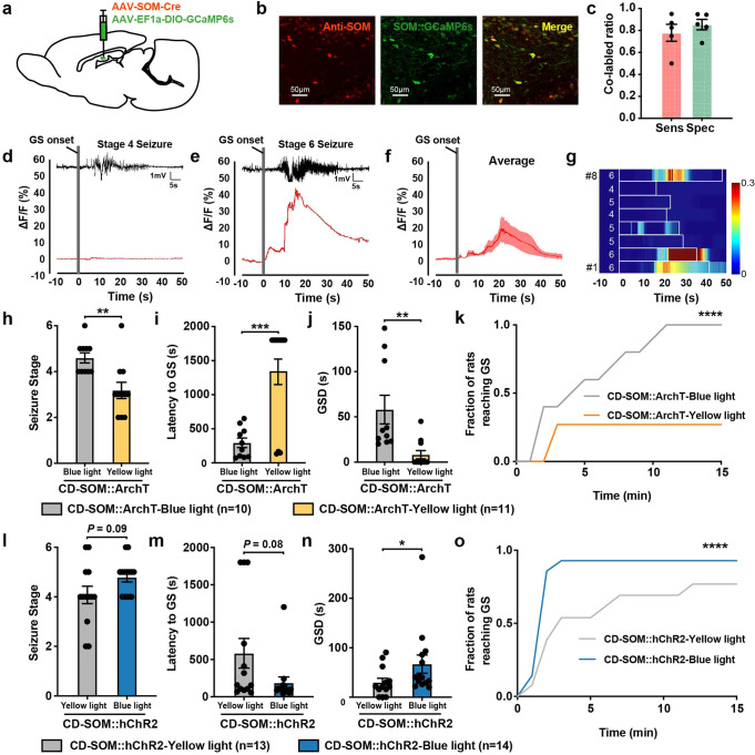
Why do seizures spread from a focal abnormality into a broader network? In cortical dysplasia, the cortical lesion alone may not tell the whole story. A study published in Signal Transduction and Targeted Therapy on May 17, 2023 linked patient recordings with a rat model and examined the distant hippocampus. Somatostatin-positive interneurons, usually associated with inhibition, were recruited during seizure spread; increasing their activity prolonged generalized seizures in the model. Different interneurons can thus contribute differently within the same pathological network.
Patient data link hippocampal epileptogenicity with network extent in a subset of cortical dysplasia. In rats, a dentate SOM-related population promotes seizure spread while a PV-related population retains inhibitory effects. Enhanced glutamatergic transmission provides a mechanistic lead.
Contents · 7 sections
A distant hippocampus can join the seizure network
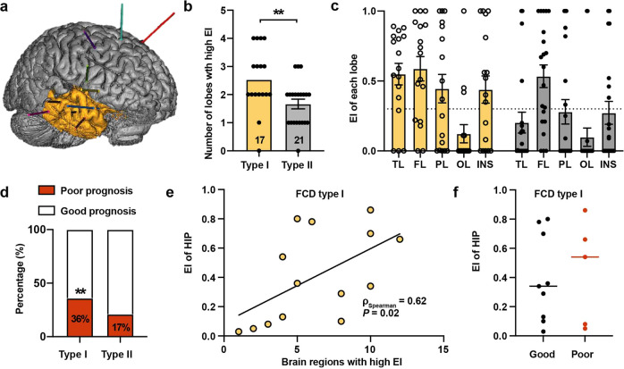
The analysis included 38 patients undergoing presurgical stereoelectroencephalography with subsequent pathological confirmation of focal cortical dysplasia. Type I cases had a broader distribution of highly epileptogenic regions. Within type I, hippocampal epileptogenicity correlated with the number of highly epileptogenic regions.
This clinical association does not establish the hippocampus as the cause of spread. Among type I patients with follow-up, hippocampal values were numerically higher with poorer outcomes, but the comparison was not significant. Network association and individual surgical outcome therefore remain distinct questions.
Source: Zheng Y et al. · Signal transduction and targeted therapy · CC BY
Original figure and English caption ↗
Dentate hyperexcitability emerges in the rat model
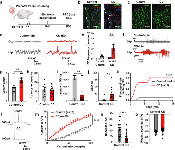
The rat model displayed cortical disorganization and abnormal EEG activity resembling clinical features. Hippocampal interictal discharges were more frequent, and provoked seizures generalized more readily and lasted longer, motivating a closer examination of hippocampal cells.
Dentate granule cells required less input to fire and produced more spikes, while resting potential and selected other properties did not significantly change. Comparable excitability changes were not found in CA1 or subicular pyramidal cells, indicating regional and cellular differences.
Source: Zheng Y et al. · Signal transduction and targeted therapy · CC BY
Original figure and English caption ↗
Fewer cells need not mean a weaker functional contribution
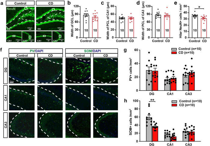
SOM-positive cell density was reduced in the dentate hilus, whereas PV-positive density did not significantly change. Granule-cell and CA1/CA3 pyramidal-layer widths were also comparable, showing distinct changes among neighboring cellular measures.
Reduced SOM-cell density appeared early in development, while limited apoptosis labeling did not resolve the full process. The remaining population’s contribution during seizures therefore became important: cell number, activity and network effect need not move in the same direction.
Source: Zheng Y et al. · Signal transduction and targeted therapy · CC BY
Original figure and English caption ↗
A SOM-related population contributes to generalized seizures
SOM-related calcium rose during generalized seizures, particularly at higher severity. Inhibition reduced stage, delayed generalization, shortened generalized-seizure duration and changed progression, supporting a contribution to seizure spread in the model.
Activation increased generalized-seizure duration and shifted cumulative progression toward faster spread. Individual stage and generalization-latency comparisons were not significant, so effects were clearest in duration and overall progression rather than every measure.
Source: Zheng Y et al. · Signal transduction and targeted therapy · CC BY
Original figure and English caption ↗
The PV-related population retains an inhibitory contribution
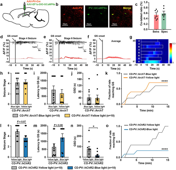
PV-related calcium responses were weaker during seizures, and functional effects differed from SOM. Activation shortened generalized-seizure duration and slowed cumulative progression, while inhibition accelerated cumulative progression. Not all individual stage or timing measures changed significantly.
Source: Zheng Y et al. · Signal transduction and targeted therapy · CC BY
Original figure and English caption ↗
Glutamatergic transmission offers an explanation
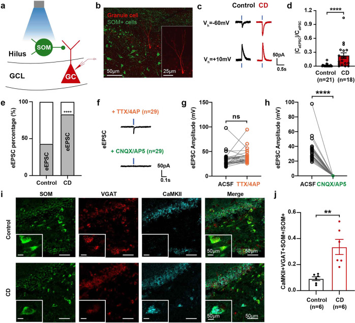
When the SOM-related population was activated, granule cells in the model more often showed excitatory currents and a higher excitatory-to-inhibitory balance. Receptor-blockade evidence supported direct glutamatergic transmission, and tissue labeling found mixed excitatory/inhibitory markers in a subset of SOM cells.
These findings support altered transmitter characteristics in a subset, without pairwise proof of co-release from every individual cell or a claim that all SOM cells become excitatory.
Source: Zheng Y et al. · Signal transduction and targeted therapy · CC BY
Original figure and English caption ↗
Understanding seizure spread reaches beyond the lesion
The study suggests that distant hippocampal cellular changes may contribute to widespread activity associated with cortical lesions. Patient network associations and rat cellular mechanisms are complementary levels of evidence. Equivalent transmitter changes in human SOM cells and their therapeutic relevance still require direct clinical evidence.