When astrocytic mitochondria lose balance, movement changes
A mouse study connects mitochondrial state in nigral astrocytes with transmitter accumulation, reversible turning and persistent motor changes after repeated perturbation, revealing the importance of neuronal support systems.
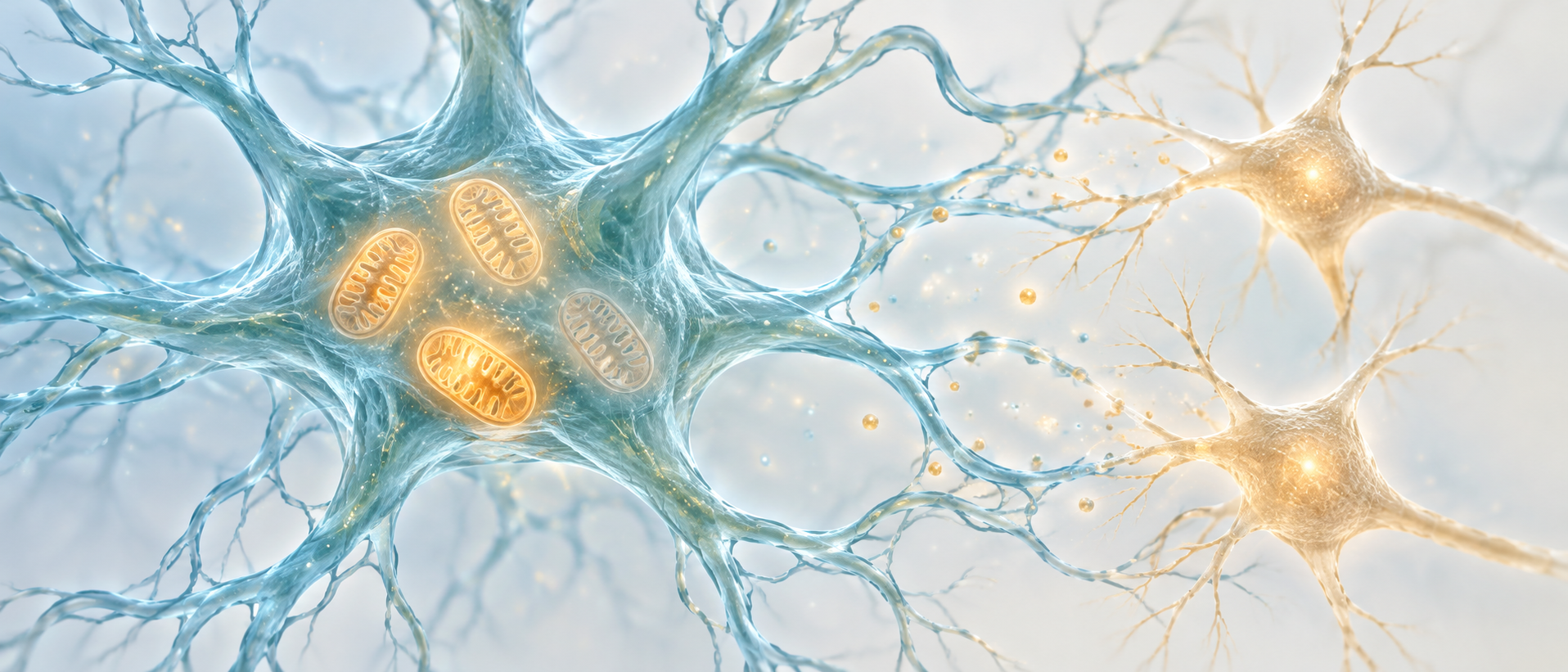
Parkinson’s disease often brings to mind the loss of nigral dopamine neurons. These cells do not work alone: neighboring astrocytes help maintain energy supply and neurotransmitter balance. A 2024 MedComm study moves to a smaller scale—the mitochondria inside astrocytes. In adult male mice, perturbing their mitochondrial membrane potential in substantia nigra pars compacta first alters local transmitter signals and turning behavior. Repeated perturbation produces persistent motor and tissue changes.
Brief astrocytic mitochondrial depolarization accompanies increased GABA and glutamate signals, reduced nigral neuronal calcium signals and reversible turning. Repeated perturbation produces slower movement, fewer TH-positive cells and Parkinson’s-disease-like features.
Contents · 7 sections
An energy station within the support system
Astrocytes support neurons and help process surrounding transmitters. Their mitochondria provide more than energy: associated metabolic enzymes also work there. The team asks whether imbalance inside support cells can alter movement without directly injuring neurons at the outset.
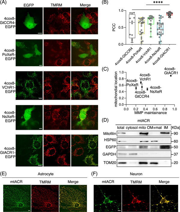
The study uses mtACR, a light-responsive tool that alters mitochondrial membrane potential. Cellular images and localization analyses show mitochondrial targeting, with potential maintained before activation in the tested cultures. This enables comparison before and after perturbation, connecting mitochondrial state itself with subsequent neural activity and behavior.
Source: Li SM et al. · MedComm · CC BY
Original figure and English caption ↗
A potential change accompanies a lower energy signal
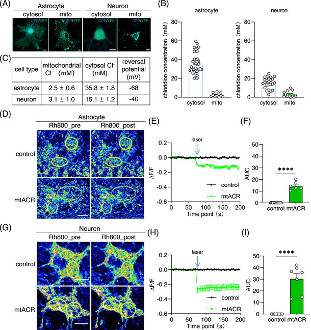
The voltage difference across the mitochondrial inner membrane helps sustain its work. After mtACR activation, potential-sensitive fluorescence decreases, supporting mitochondrial depolarization. Cultured neurons and astrocytes both show this change, with different amplitudes.
The ATP indicator signal also decreases in cultured astrocytes, whereas the tested reactive-oxygen signal does not significantly increase. Energy and membrane-potential measures change without a parallel worsening of every mitochondrial readout.
Source: Li SM et al. · MedComm · CC BY
Original figure and English caption ↗
Turning changes before overall movement declines
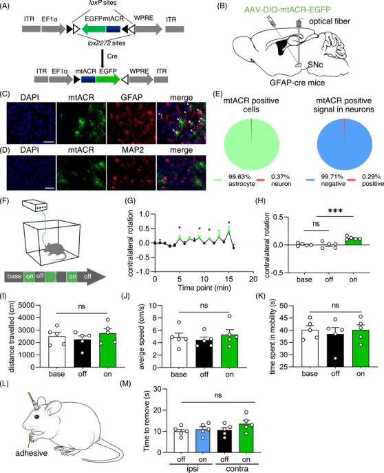
Brief perturbation of astrocytic mitochondria on one side of the mouse substantia nigra pars compacta increases turning toward the opposite side. Turning diminishes when perturbation ends. Distance traveled, average speed, time moving and forepaw adhesive-removal time do not significantly change.
Movement is not broadly slowed at this stage. TH staining intensity and tested glial counts also do not significantly change under brief perturbation. A functional shift emerges before these tissue measures decline, suggesting altered local cellular interactions.
Source: Li SM et al. · MedComm · CC BY
Original figure and English caption ↗
Two transmitters rise while the network shifts toward inhibition
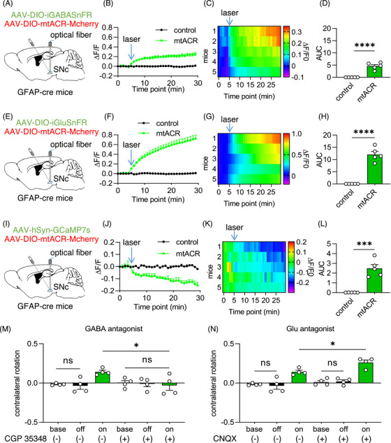
Local optical recordings show increased GABA and glutamate sensor signals but decreased population calcium signals in nigral neurons. These transmitters are commonly linked with inhibition and excitation, yet their increases do not simply cancel at the network level.
Pharmacological interventions further separate their contributions. GABA-receptor blockade reduces turning, whereas blockade of the tested glutamate receptors worsens it. GABA action therefore has an important contribution under these conditions. Signal amplitudes from different sensors also depend on their respective sensitivities.
Source: Li SM et al. · MedComm · CC BY
Original figure and English caption ↗
Repeated perturbation leaves persistent motor changes
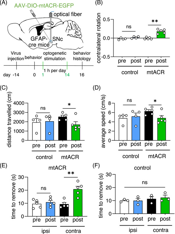
What happens when a reversible disturbance recurs? After two weeks of intermittent perturbation, mice show spontaneous contralateral turning even without current tool activation. Distance traveled and average speed decrease, and adhesive removal from the contralateral forepaw takes longer.
Gross and fine motor measures that did not significantly change during brief perturbation now differ. Controls do not show the same changes. Persistent abnormalities extend the story from immediate network imbalance to longer-term cellular support.
Source: Li SM et al. · MedComm · CC BY
Original figure and English caption ↗
Tissue changes accompany the motor phenotype
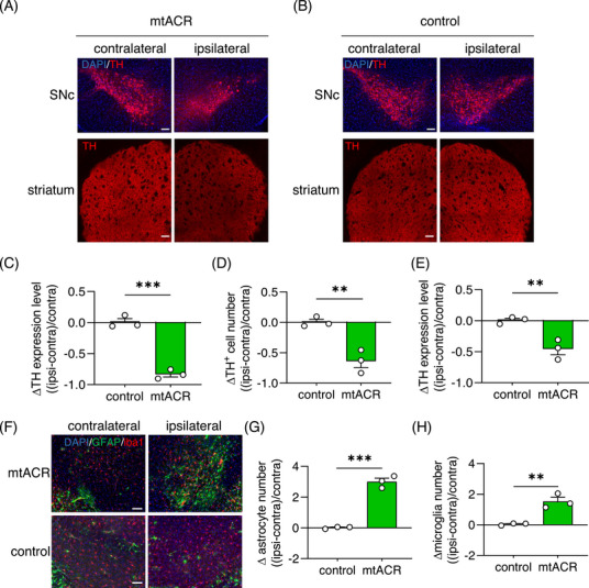
In tissue, TH staining decreases in the perturbed nigra and striatum, with fewer TH-positive cells in nigra. Supplementary results show alpha-synuclein aggregation and increased GABA. Counts of labeled astrocytes and microglia increase in the sampled region.
Together with persistent motor deficits, these changes constitute Parkinson’s-disease-like features. A perturbation beginning inside astrocytes extends to transmitters, neuronal markers and movement, showing how strongly support-system state can affect the neuronal environment.
Source: Li SM et al. · MedComm · CC BY
Original figure and English caption ↗
Motor circuits depend on surrounding metabolic balance
The study connects mitochondrial state, transmitter handling and movement. Protein levels of the GABA-metabolism enzyme ABAT decrease, whereas the change in the related enzyme GDH is nonsignificant. Metabolic processes may contribute to transmitter accumulation, with their precise steps still to be distinguished.
This is an induced-perturbation study in adult male mice, revealing one condition that produces disease-like features. Its concrete insight is that movement depends on neurons and also on energy state within support cells and the transmitter balance they maintain around the network.