How can motor-cortex activity contribute after spinal cord injury?
In rats, increasing activity in excitatory-associated M1 cells is followed by partial improvements in movement and motor-evoked responses, alongside differences in lesion cavities and growth-related markers.
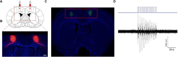
Spinal cord injury disrupts communication between brain and body. Beyond changes at the lesion, what can the brain’s motor-command system contribute to recovery? A rat study published in Frontiers in Neuroscience on April 9, 2021 increases activity in excitatory-associated cells of primary motor cortex (M1). Later hindlimb performance and motor-evoked responses improve, alongside differences in lesion tissue and molecular markers. The findings extend attention toward brain–spinal interactions while leaving open how the changes connect.
Increasing activity in the targeted M1 population is followed by partial recovery of hindlimb function and motor-evoked responses, smaller spinal cavities and higher BDNF, NGF and growth-related markers. These accompanying changes do not yet establish the complete repair mechanism.
Contents · 7 sections
Looking for a contribution from the brain
M1 contributes to translating movement plans into descending commands. Brain activity and connections can reorganize after injury. Earlier observations linked motor-cortex excitability with later recovery, but regional activity alone does not identify which of its diverse cells contribute.
This study primarily targets excitatory M1 cells and examines the effects of increasing their activity. Recordings show increased firing in sampled cells with accompanying hindlimb movement. The subsequent question is whether injured rats show better function during follow-up.
Source: Deng WW et al. · Frontiers in neuroscience · CC BY
Original figure and English caption ↗
Movement improves, with recovery still incomplete
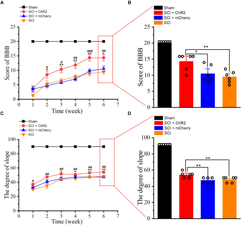
All injured groups improve during follow-up, with greater improvement in the targeted-activation group. At six weeks, hindlimb locomotor scores exceed both the injury-only and fluorescent-control groups, alongside better stability on an inclined surface. The tests provide complementary views of motor function.
The two injured control groups do not differ significantly, while the targeted-activation group still remains below the uninjured control. The result is partial improvement in injury-related function rather than complete restoration of the motor system.
Source: Deng WW et al. · Frontiers in neuroscience · CC BY
Original figure and English caption ↗
Motor-evoked responses provide a second perspective
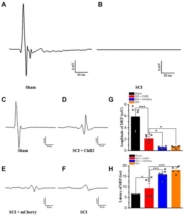
Beyond behavior, the study examines motor-evoked potentials. Responses are almost absent early after injury. At follow-up, the targeted-activation group shows larger responses and shorter latency than both injured controls.
A motor-evoked potential combines the state of a long motor pathway into a comparable response: amplitude reflects the strength of combined activity, while latency describes how quickly the response appears. Stronger, faster responses fit the hindlimb improvement, but still fall short of the uninjured group.
Source: Deng WW et al. · Frontiers in neuroscience · CC BY
Original figure and English caption ↗
Lesion tissue also differs
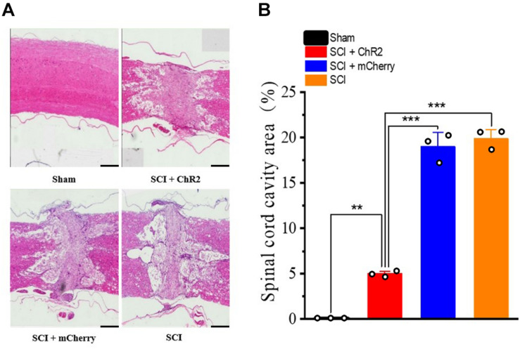
At six weeks, the targeted-activation group has a smaller cavity area at the spinal lesion than both injured controls. The difference extends beyond behavior and electrical responses to tissue structure, linking increased brain activity with a different state at the distant lesion.
These endpoint sections compare final tissue states across groups. Smaller cavities accompany better function, while the relative contributions of tissue preservation, later remodeling and other repair processes remain to be tracked over time.
Source: Deng WW et al. · Frontiers in neuroscience · CC BY
Original figure and English caption ↗
Growth-related proteins accompany tissue differences
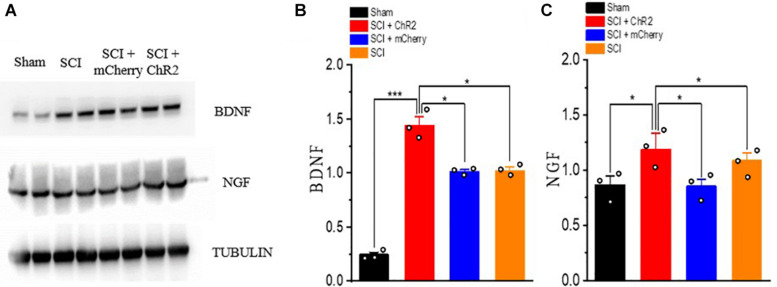
In earlier lesion-tissue protein measurements, BDNF and NGF levels are higher in the targeted-activation group than in both injured controls. These neurotrophic proteins are associated with cell survival and plasticity, offering candidate links between brain activity and spinal tissue changes.
Repair involves not only descending transmission but also the growth environment around the lesion. Higher BDNF and NGF offer a possible link between these levels. They accompany recovery here, while their status as required intermediates remains unresolved.
Source: Deng WW et al. · Frontiers in neuroscience · CC BY
Original figure and English caption ↗
Stronger fiber-related labeling leaves reconstruction to be resolved
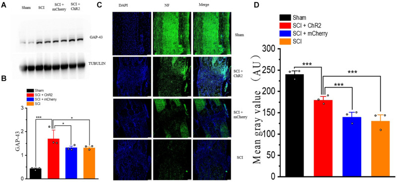
GAP-43, associated with axonal growth and synaptic plasticity, is also higher in the targeted-activation group. At six weeks, lesion-region neurofilament fluorescence exceeds both injured controls but remains below the uninjured group. Together, these markers indicate differences in fiber-associated tissue state.
Spared fibers, axonal sprouting and new growth can each strengthen fiber-related labeling. The fluorescence is therefore a snapshot of tissue state. Determining which fibers cross the lesion and reconnect motor pathways requires following those connections more directly.
Source: Deng WW et al. · Frontiers in neuroscience · CC BY
Original figure and English caption ↗
Putting brain and spinal cord into the same recovery picture
The rat study connects M1-related activity with movement, evoked transmission and lesion-tissue differences, placing brain–spinal interactions within recovery. The findings converge across levels, while the complete causal sequence remains unresolved.
The study does not directly compare the intervention with electrical or magnetic stimulation, or separately test inhibitory-cell contributions. It provides animal evidence at the population level for understanding how motor cortex can influence recovery after spinal cord injury.