How an active prefrontal cortex supports repair after white-matter injury
Prefrontal activation improves working memory and callosal myelin-related measures in male mice with chronic hypoperfusion. Mature oligodendrocytes increase, and Wnt2 experiments offer a candidate mechanism.
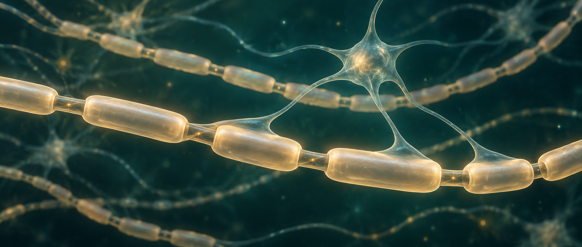
Fast neural signaling depends on both neuronal activity and myelin around axons. After chronic hypoperfusion injures white matter, how does this wrapping relate to memory? A study published online in Advanced Science on December 18, 2022 activates glutamatergic-related medial prefrontal (mPFC) cells in a mouse bilateral-carotid-stenosis model. Following axons through corpus callosum, the team observes improvements in working memory, myelin structure and oligodendrocyte maturation, then investigates Wnt2 signaling. Neurons may influence not only communication along existing routes but also the surrounding cellular wrapping.
Prefrontal activation accompanies myelin repair, maturation and improved working memory. Wnt2 increases, overexpression and culture tests further connect neuronal activity with myelin-cell maturation.
Contents · 6 sections
Memory changes accompany white-matter injury
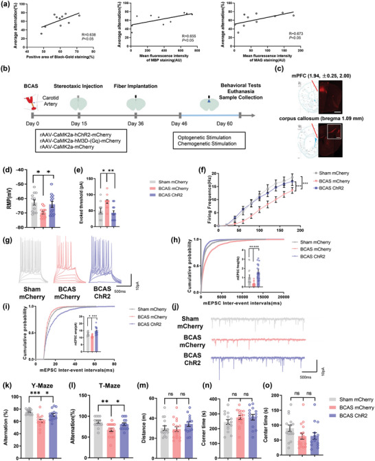
After model establishment, callosal myelin staining and related protein measures correlate with maze performance. Superficial prefrontal projections traverse corpus callosum, motivating the target. Repeated activation improves Y- and T-maze alternation without significant changes in open-field distance or center and corner time.
Maze alternation tests use of recently encountered information, principally reflecting short-term working memory. Model mice show lower cellular excitability and miniature excitatory synaptic currents, which improve after activation. Behavioral changes thus accompany cellular functional differences without a general increase in locomotion.
Source: Deng S et al. · Advanced science (Weinheim, Baden-Wurttemberg, Germany) · CC BY
Original figure and English caption ↗
Wrapping around axons shows structural improvement
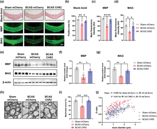
The corpus callosum connects the cerebral hemispheres and supports communication between them. Callosal Black-gold staining shows reduced myelin signal in model mice and improvement after prefrontal activation. Staining and protein measurements of MBP and MAG also increase, providing multiple tissue-level views alongside behavior.
Electron microscopy shows a higher proportion of myelinated axons and a lower g-ratio, a measure of relative wrapping thickness. These structural findings support improved myelin status through convergent observations spanning protein signals and axonal wrapping.
Source: Deng S et al. · Advanced science (Weinheim, Baden-Wurttemberg, Germany) · CC BY
Original figure and English caption ↗
Another activation approach links tissue and conduction measures
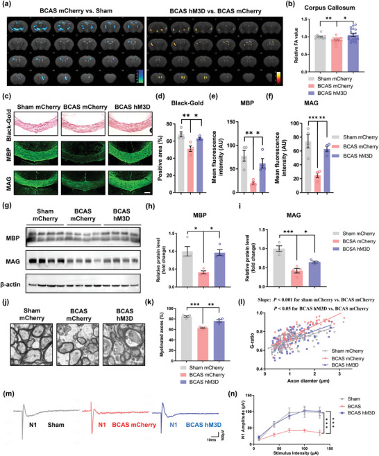
Chemogenetic activation produces corresponding working-memory and tissue improvements. Callosal fractional anisotropy decreases in model mice and rises after activation, alongside staining, protein and ultrastructural changes. Imaging and tissue observations jointly characterize white-matter status.
In callosal slice compound-action-potential recordings, the early peak associated with fast myelinated conduction increases in amplitude. This offers a functional observation beyond structure. This connects structural observations of myelin with a measurable pathway response.
Source: Deng S et al. · Advanced science (Weinheim, Baden-Wurttemberg, Germany) · CC BY
Original figure and English caption ↗
More cells mature without a parallel proliferation increase
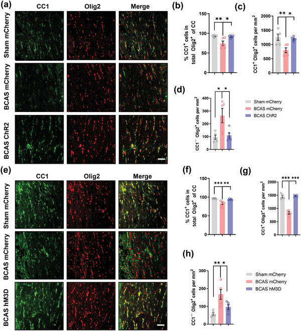
Oligodendrocytes produce myelin. Lineage and maturation markers show lower mature-cell proportion and density in model mice, with increases after prefrontal activation and a decrease in immature-related density. Both activation approaches show this direction.
Proliferation markers, selected newly labeled-cell counts and death markers show little change, while maturation among newly labeled cells increases. The pattern focuses on maturation rather than greater proliferation alone. Total lineage-cell numbers also increase, prompting a migration hypothesis without direct migration tracking.
Source: Deng S et al. · Advanced science (Weinheim, Baden-Wurttemberg, Germany) · CC BY
Original figure and English caption ↗
Wnt2 emerges between neurons and myelin-producing cells
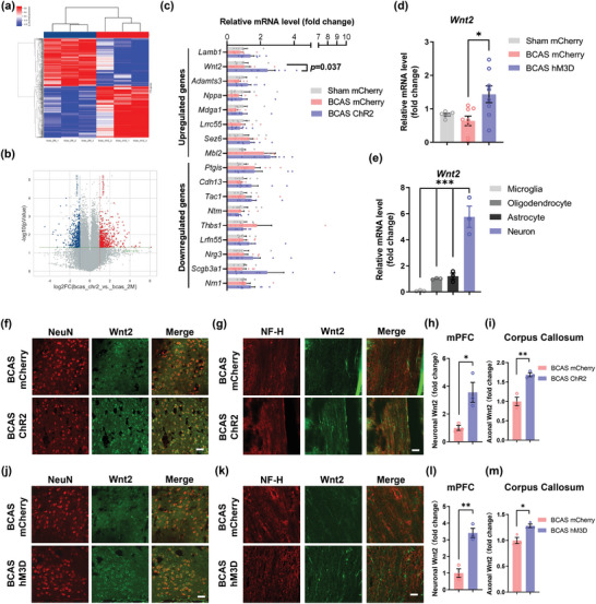
Expression screening and validation of candidate secreted-related molecules identify increased Wnt2 with both activation approaches. Staining shows more related protein in prefrontal cortex and corpus callosum, partly distributed along axons. Wnt-related beta-catenin expression also increases in oligodendrocyte-lineage cells.
These findings identify Wnt2 as a candidate bridge through which active neurons may influence nearby myelin cells. Axonal protein distribution supports the idea without directly recording release timing or quantity. The team next tests whether increased Wnt2 expression can promote similar changes.
Source: Deng S et al. · Advanced science (Weinheim, Baden-Wurttemberg, Germany) · CC BY
Original figure and English caption ↗
Increasing Wnt2 reproduces a repair-related pattern
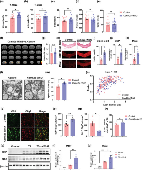
Increasing prefrontal Wnt2 in model mice improves working memory, diffusion-imaging and myelin-structure measures and increases mature oligodendrocytes, with largely unchanged open-field locomotion and zone times. In cultures with an existing differentiation stimulus, added Wnt2 increases related myelin proteins, and Wnt-signaling antagonism weakens that enhancement.
Overexpression and culture findings support promotion by Wnt2 without testing whether in-vivo Wnt2 blockade eliminates prefrontal-activation benefits. In male mice entering the study at eight weeks, neural activity and myelin-cell maturation are linked after chronic hypoperfusion. Older animals and a fuller causal chain remain further questions.
Source: Deng S et al. · Advanced science (Weinheim, Baden-Wurttemberg, Germany) · CC BY
Original figure and English caption ↗