After peripheral nerve injury, the cerebellum also shapes motor recovery
In mice, abnormal cerebellar circuitry slows motor recovery after peripheral nerve injury. Modifying deep-nuclear activity or increasing Gria1 expression improves measured functions, with a small human sample providing associative evidence.
After a peripheral nerve is injured, does recovery depend only on reconnecting interrupted communication? Muscle reinnervation matters, but central organization of movement may also shape the outcome. A study published in npj Regenerative Medicine on September 5, 2022 examines sciatic-nerve injury alongside cerebellar function in mice. Despite similar measured sensory recovery, mice with abnormal cerebellar circuits show poorer grip-strength and muscle-response recovery. Following deep cerebellar nuclei, DCN, and glutamate-receptor regulation links recovery after peripheral injury with central-network function.
Cerebellar abnormalities affect post-injury motor recovery. DCN stimulation, baclofen-related modulation and pre-injury Gria1 overexpression each improve measured recovery. Human case and expression data support associations without testing these interventions in people.
Contents · 7 sections
Similar injury, different recovery trajectories
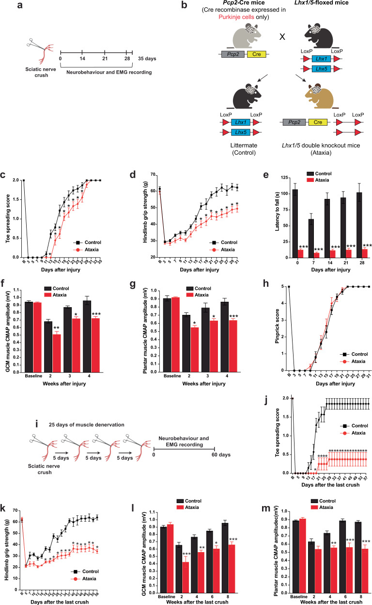
The study uses ataxic mice with conditional Lhx1/Lhx5 deletion in Purkinje cells. Before injury, coordination is poorer but measured grip strength, toe spreading and muscle-response baselines resemble controls. After sciatic-nerve injury, controls recover while grip strength and compound muscle action potentials remain below baseline in ataxic mice.
Pinprick recovery and measured sensory-axon growth show no corresponding difference. Longer muscle denervation widens the motor-recovery gap. Similar peripheral injury does not guarantee the same subsequent trajectory of movement.
Source: Asthana P et al. · NPJ Regenerative medicine · CC BY
Original figure and English caption ↗
Not every brain lesion produces the same difference
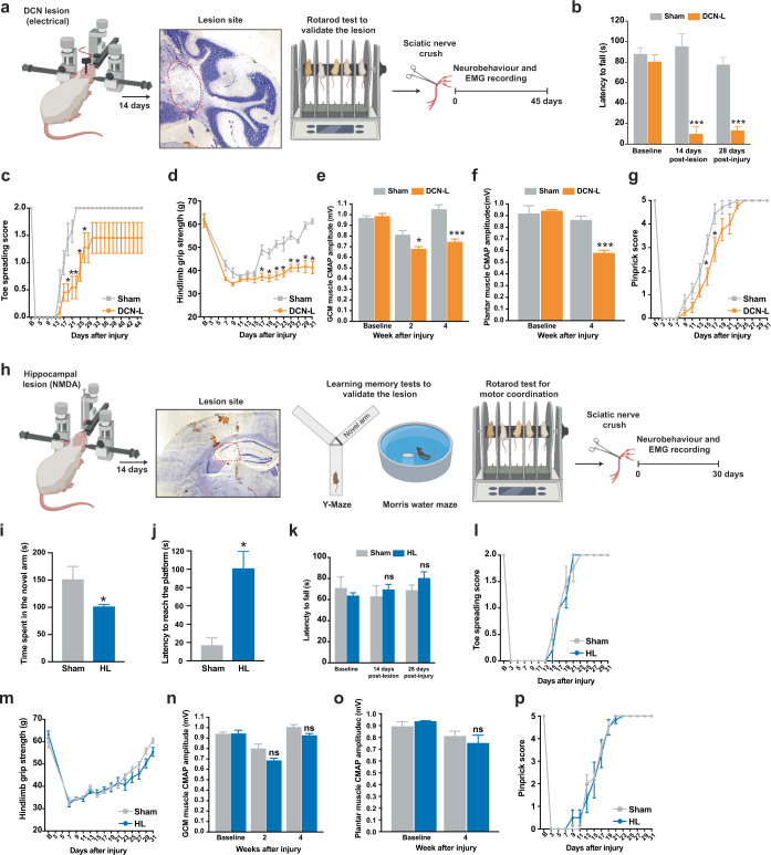
DCN lesions also impair toe-spreading, grip and muscle-response recovery, with only a brief sensory delay. Hippocampal lesions impair spatial tasks, yet measured motor and sensory functions still recover after peripheral injury.
The comparison focuses attention on motor-related circuitry. Purkinje-cell inhibition of deep nuclei helps fine-tune motor output; abnormal regulation can affect functional rebuilding after injury. The hippocampal comparison concerns these conditions rather than excluding every other region.
Source: Asthana P et al. · NPJ Regenerative medicine · CC BY
Original figure and English caption ↗
Responses from central stimulation become slower and weaker
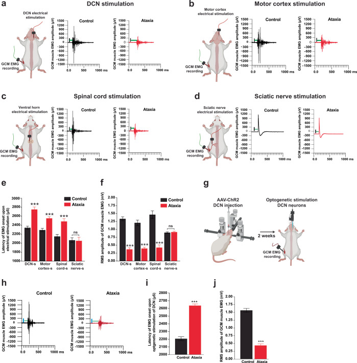
Stimulating DCN, motor cortex or spinal cord produces later and smaller muscle responses in ataxic mice. Direct sciatic-nerve stimulation yields similar measured response amplitude and conduction timing to controls. The difference therefore extends beyond peripheral signal transmission alone.
Optogenetic DCN activation likewise evokes slower, smaller muscle responses. Converging stimulation results connect abnormal cerebellar output with descending motor-system function, using muscle activity as an endpoint.
Source: Asthana P et al. · NPJ Regenerative medicine · CC BY
Original figure and English caption ↗
Modifying abnormal firing accompanies better recovery
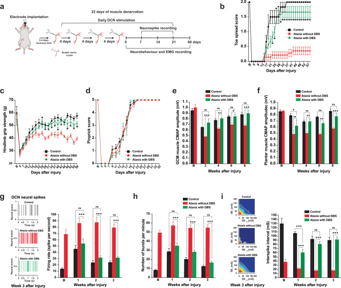
DCN recordings show higher mean firing and more bursts in ataxic mice. Nuclear electrical stimulation reduces these abnormalities. Greater excitation does not automatically mean better motor recovery; the organization of firing also matters.
With prolonged muscle denervation, stimulated mice recover toe spreading, grip and muscle responses toward control levels, while unstimulated mice retain deficits. Measured sensory recovery shows no corresponding group difference. The findings link activity modulation with improved motor function.
Source: Asthana P et al. · NPJ Regenerative medicine · CC BY
Original figure and English caption ↗
Metabolite changes point toward the glutamate system
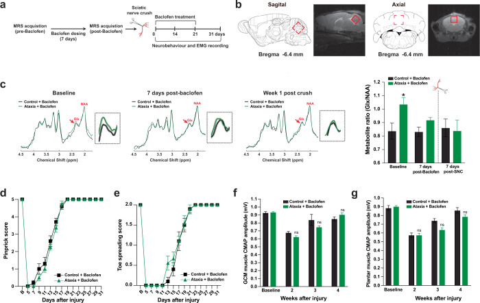
Magnetic resonance spectroscopy detects a higher combined glutamate/glutamine signal, Glx, in ataxic cerebellum. This reflects regional metabolite levels rather than momentary synaptic glutamate release.
After the GABA-receptor-related drug baclofen, Glx approaches control levels. Nerve injury follows confirmation of this change, with better functional and muscle-response recovery; separate recordings also show improved firing patterns. This leads to examination of receptor-level changes.
Source: Asthana P et al. · NPJ Regenerative medicine · CC BY
Original figure and English caption ↗
Gria1 expression adds another line of evidence
After injury, DCN Gria1 transcripts rise in controls but not in ataxic mice. Gria1 encodes the GluA1 subunit of AMPA-type glutamate receptors. Its divergent response stands out among changes in several receptor genes.
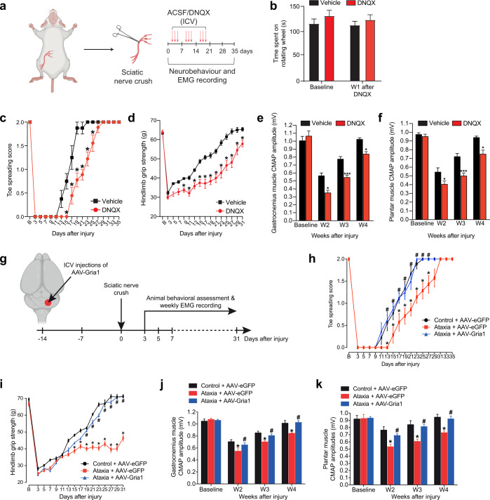
DNQX blockade of AMPA-related transmission slows normal-mouse motor recovery without significant changes in baseline rotarod or measured sensory recovery. Conversely, increasing DCN Gria1 before injury improves toe spreading, grip and muscle-response recovery in ataxic mice. Pharmacological blockade and gene enhancement complement each other, while DNQX is not selective for GluA1.
Source: Asthana P et al. · NPJ Regenerative medicine · CC BY
Original figure and English caption ↗
Human associations accompany mouse mechanistic evidence
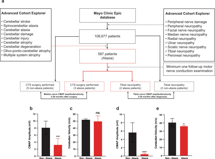
Public expression datasets show lower GRIA1 in some ataxia-related tissues or cells, alongside several nonsignificant comparisons. A retrospective clinical analysis ultimately includes twelve patients. Ataxic cases have lower muscle-response amplitudes after carpal-tunnel surgery or during tibial-neuropathy follow-up, while wrist motor-conduction velocity differs nonsignificantly.
The small case sample and mouse interventions raise a shared question: can central motor networks provide suitable support during peripheral recovery? Mechanistic results obtained mainly in adult male mice extend attention from the injured nerve to cerebellar circuits. The human analyses do not test stimulation, drug or gene interventions.
Source: Asthana P et al. · NPJ Regenerative medicine · CC BY
Original figure and English caption ↗