Abnormal thalamic synchrony reveals a clue to absence seizures
In BK-channel mutant mice, midline-thalamic cells burst synchronously during absence seizures. Changing firing patterns, increasing behavioral engagement or applying closed-loop stimulation reduces tested spike-wave episodes and behavioral arrest.
A mouse suddenly stops moving as repeated spike-wave discharges appear in its EEG. Such absence-like episodes occur frequently in mice carrying the BK-channel D434G mutation. A 2024 PNAS study follows the timing of EEG and cellular activity to the midline thalamus, where many cells burst together during seizures. Changing their firing pattern reduces abnormal EEG activity and behavioral arrest, adding a circuit component to the mechanism in this model.
During absence seizures, midline-thalamic cells reduce mean firing while synchronized bursting increases. Reducing bursting or changing firing patterns suppresses tested episodes in BK-D434G mice.
Contents · 7 sections
The midline thalamus shows stronger activity labeling
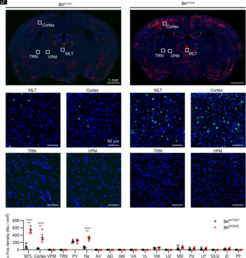
BK is a large-conductance potassium channel involved in cellular electrical activity. The gain-of-function D434G mutation produces frequent spike-wave episodes and behavioral arrest in this mouse model. Mutants show more c-Fos activity labeling in cortex and midline thalamus (MLT) than controls.
Thalamic regions do not all look alike. Several MLT nuclei stand out, whereas thalamic reticular and ventral posteromedial nuclei lack comparable labeling increases. Because c-Fos reflects both firing strength and pattern, the team next records spikes to determine the activity underlying the labeling.
Source: Dong P et al. · Proceedings of the National Academy of Sciences of the United States of America · CC BY-NC-ND
Original figure and English caption ↗
Less firing becomes more synchronized
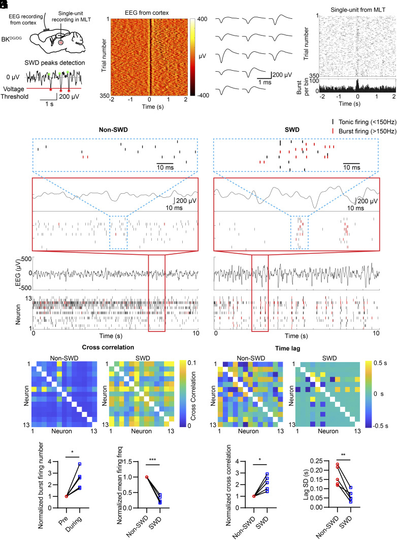
Aligning cellular spikes with EEG reveals the change during seizures: MLT mean firing falls, but bursting increases around spike-wave peaks. Different cells more often emit rapid spike groups at nearly the same time, with smaller timing differences.
Stronger activity labeling can therefore coexist with a lower mean firing rate. The network resembles many players switching to tightly coordinated short phrases. Synchrony and spike arrangement provide information beyond a single average.
Source: Dong P et al. · Proceedings of the National Academy of Sciences of the United States of America · CC BY-NC-ND
Original figure and English caption ↗
Mutant cells produce stronger rebound bursts
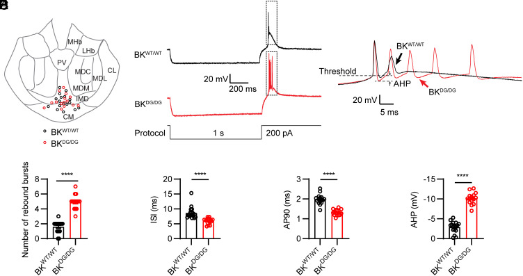
Slice recordings show stronger rebound bursts after hyperpolarization in mutant MLT cells: more spikes, shorter intervals, faster action-potential repolarization and a deeper afterhyperpolarization. These differences connect the abnormal network rhythm with intrinsic cellular properties.
T-type calcium channels contribute to thalamic rebound bursts, while BK channels influence repolarization. The authors propose that enhanced BK function may help cells regain readiness to spike more rapidly, strengthening bursts. This model connects channel changes with firing rhythm.
Source: Dong P et al. · Proceedings of the National Academy of Sciences of the United States of America · CC BY-NC-ND
Original figure and English caption ↗
Spike-wave episodes decline when movement begins
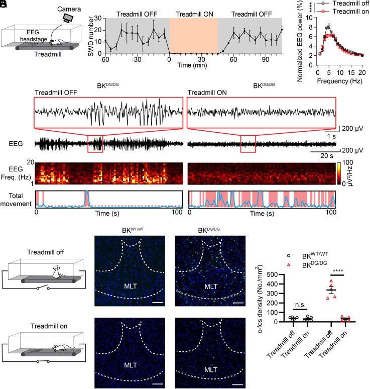
On a stationary treadmill, mice develop frequent spike-wave episodes and arrest. When the treadmill moves and mice walk, spike-wave activity declines rapidly; after movement stops, abnormal activity gradually returns. Episodes are closely associated with the current behavioral state.
Movement also reduces MLT c-Fos labeling in mutants, without a significant corresponding change in controls. This connects behavioral state, episodes and thalamic activity as an immediate effect of the experimental condition.
Source: Dong P et al. · Proceedings of the National Academy of Sciences of the United States of America · CC BY-NC-ND
Original figure and English caption ↗
Tonic firing interrupts synchronized bursts
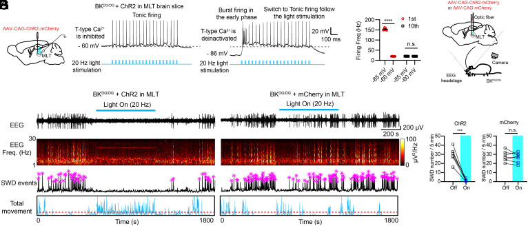
BK-channel blockade reduces MLT bursting and suppresses abnormal EEG activity, although the local anti-seizure effect wanes later. The team also drives MLT cells toward a more regular tonic mode to examine the effect of changing the firing pattern itself.
In slices, cells shift from bursting to tonic firing. In vivo, the intervention rapidly suppresses spike-wave episodes and mice move out of arrest, without a corresponding significant change in labeling controls. Increased tonic activity can thus accompany reduced abnormal synchrony.
Source: Dong P et al. · Proceedings of the National Academy of Sciences of the United States of America · CC BY-NC-ND
Original figure and English caption ↗
Behavioral activation can change thalamic rhythm indirectly
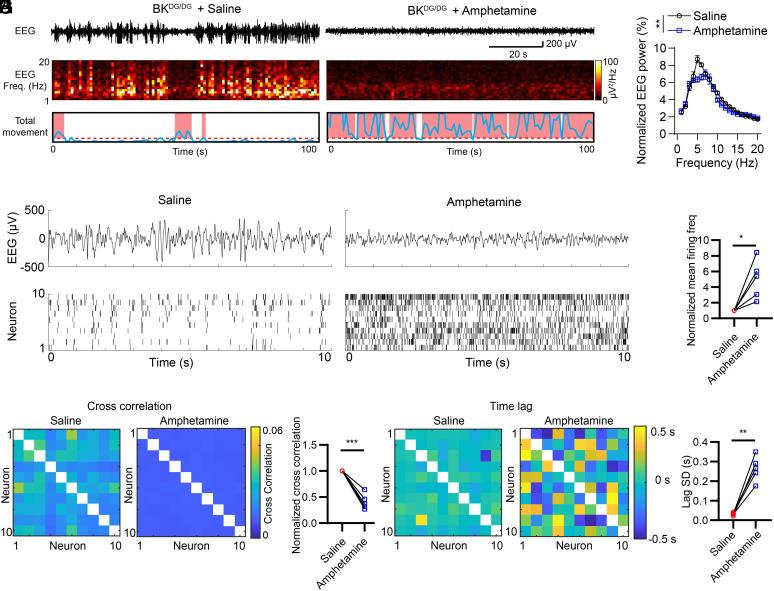
Stimulant experiments likewise increase movement, reduce spike-wave episodes and raise overall MLT firing while reducing intercellular synchrony. In slices, the drug does not directly change MLT rebound bursts, suggesting involvement of other regions in the in-vivo effect.
Direct MLT manipulation and changes through a broader network both converge on less synchronized bursting. As the network moves away from abnormal coordination, spike-wave episodes and behavioral arrest decline.
Source: Dong P et al. · Proceedings of the National Academy of Sciences of the United States of America · CC BY-NC-ND
Original figure and English caption ↗
Detecting abnormal EEG and then modulating thalamus
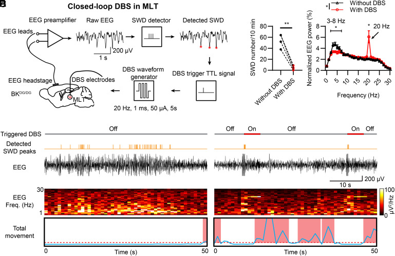
Closed-loop stimulation turns the idea into a real-time response: the system detects spike-wave events in EEG and then stimulates MLT. Episodes decline significantly and movement increases within the tested window. Without triggered stimulation, the same animal continues to show frequent episodes and arrest. Seizure-associated band power decreases while power at the stimulation frequency increases.
This BK-D434G mouse study identifies a modifiable thalamic activity pattern. Seizures combine lower mean firing with stronger synchronized bursts, while interventions can raise overall firing as abnormal EEG declines. How cells coordinate becomes central to understanding the model.
Source: Dong P et al. · Proceedings of the National Academy of Sciences of the United States of America · CC BY-NC-ND
Original figure and English caption ↗