A small pallidal ensemble behind abnormal movements
In adult mice, suppressing a pallidal ensemble recruited during dyskinesia reduces abnormal movements, while reactivation evokes them. Combined activation with a striatal ensemble produces stronger effects.
Levodopa can improve Parkinsonian motor symptoms, but prolonged treatment may be accompanied by dyskinesia: involuntary twisting and abnormal limb postures. Which cells participate in these movements? A study published in Cell Reports Medicine on May 16, 2024 labels cells active during dyskinesia in dopamine-depleted mice. The team identifies a small neuronal ensemble in external globus pallidus and uses opposing interventions to connect its activity with abnormal movements.
Suppressing external-pallidal neurons labeled during dyskinesia alleviates axial and limb abnormalities. Reactivating them in the levodopa-off state also evokes dyskinetic movements.
Contents · 7 sections
From brain-wide activity to candidate ensembles
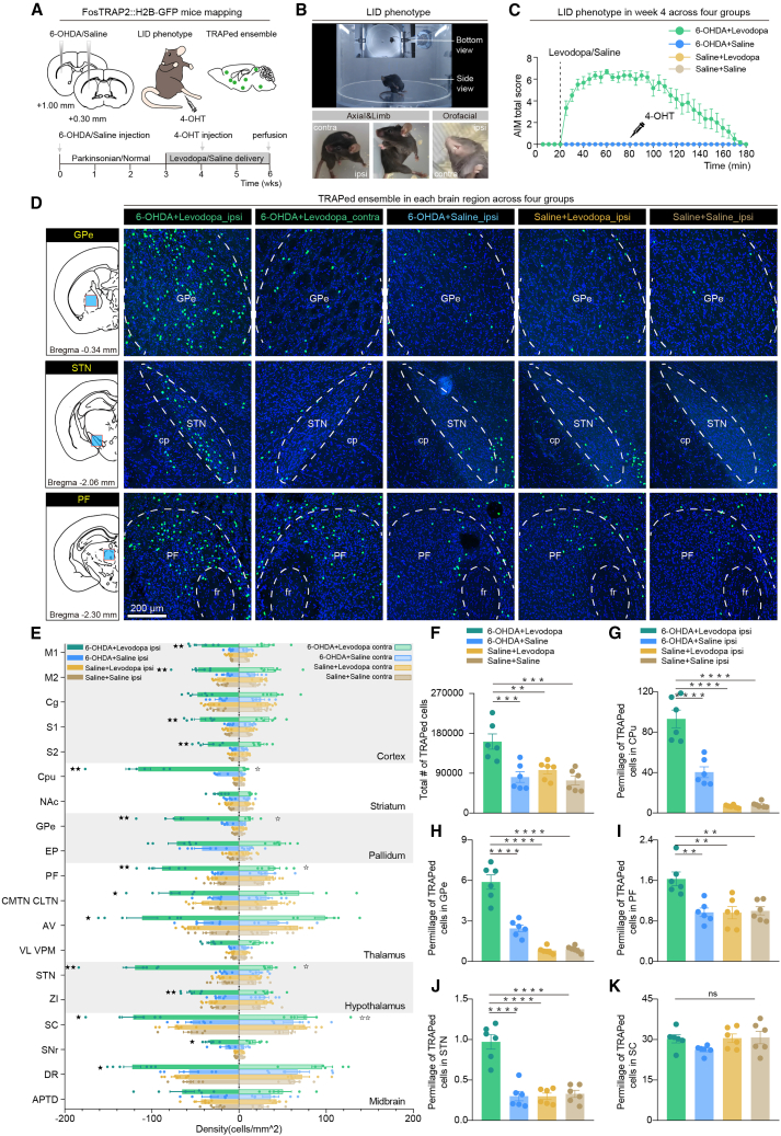
Mice develop axial, limb and orofacial abnormalities after unilateral dopaminergic lesions and repeated levodopa treatment. Activity-dependent labeling gives recruited cells an enduring identifier. Controls help distinguish changes associated with the combination of lesion, treatment and dyskinesia.
Labeled cells are widespread. Comparing groups, hemispheres and each region’s share of brain-wide labeling identifies GPe, parafascicular thalamus (PF) and subthalamic nucleus (STN), alongside the previously implicated striatum. The next question concerns which ensemble’s activity tracks dyskinesia.
Source: Shen C et al. · Cell Reports Medicine · CC BY-NC
Original figure and English caption ↗
Pallidal activity rises and falls with dyskinesia
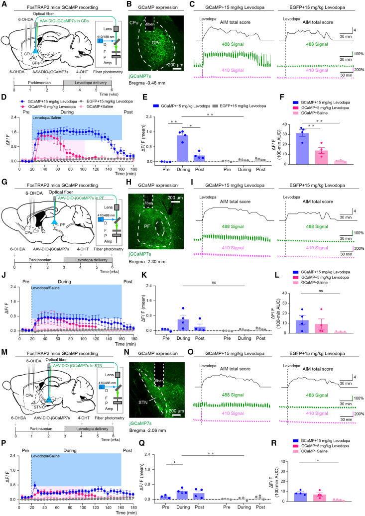
The labeled GPe ensemble shows a sustained calcium increase during a subsequent dyskinetic episode, followed by decline. More severe dyskinesia accompanies a larger signal, while milder movements accompany smaller changes. Single-cell imaging also detects increased activity.
The PF ensemble’s mean change does not reach significance. STN shows a brief onset peak rather than sustained tracking like GPe. Several regions contain labeled cells but differ dynamically, focusing further causal interventions on GPe.
Source: Shen C et al. · Cell Reports Medicine · CC BY-NC
Original figure and English caption ↗
Quieting the ensemble reduces abnormal twisting
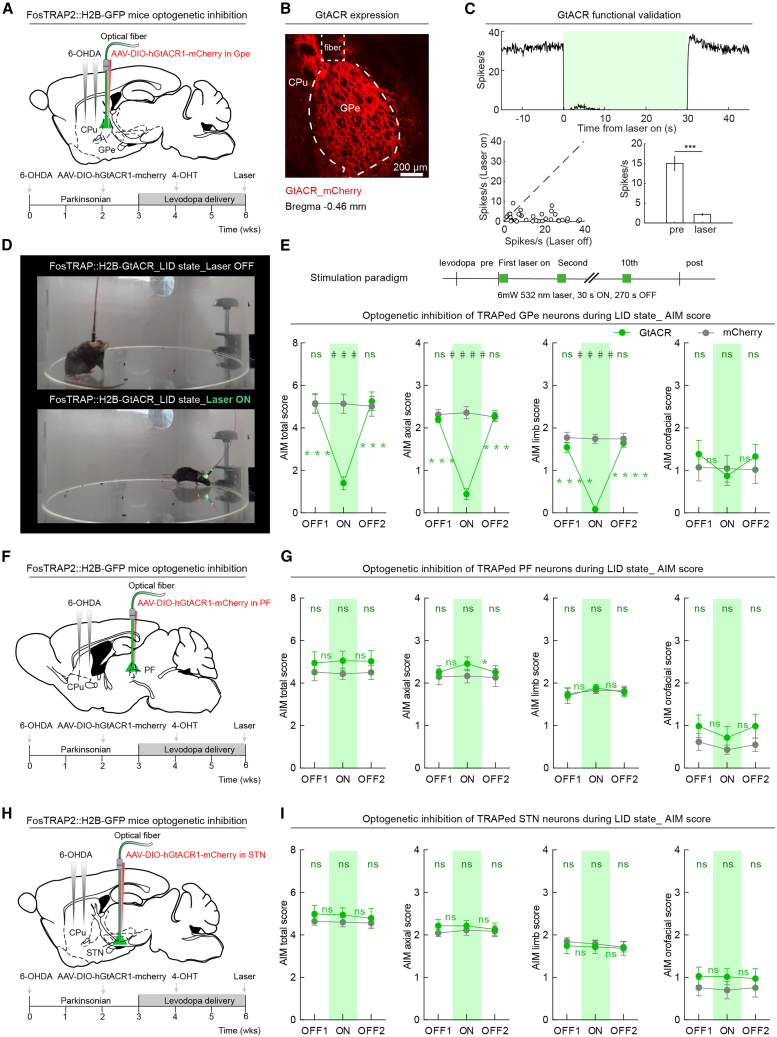
Suppressing labeled GPe neurons during pronounced dyskinesia rapidly lowers abnormal-movement scores. Axial twisting and abnormal limb postures decrease, and some mice resume straight walking and rearing. Orofacial scores do not significantly improve, showing different sensitivity across movement types.
Abnormal movements return when suppression ends, and repeated interventions still reduce them. Suppressing labeled PF or STN populations does not produce comparable overall improvement. GPe activity is thus linked to immediate changes in dyskinesia through intervention.
Source: Shen C et al. · Cell Reports Medicine · CC BY-NC
Original figure and English caption ↗
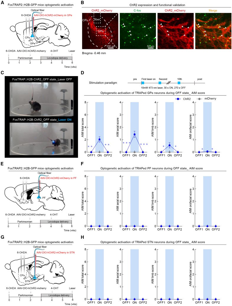
Reactivation evokes abnormalities in the levodopa-off state
The reverse experiment uses mice with this established model in the levodopa-off state. Reactivating labeled GPe cells evokes neck and trunk bending and abnormal limb postures. The movements are most prominent early in activation, then gradually transition toward voluntary movement.
Activating corresponding labeled PF or STN populations does not evoke similar abnormalities. They were recruited during dyskinesia, yet interventions differ from GPe. The distinction concerns subsets defined by their activity history rather than a single function assigned to an entire region.
Source: Shen C et al. · Cell Reports Medicine · CC BY-NC
Original figure and English caption ↗
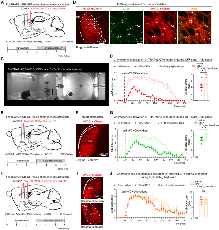
Recruiting two ensembles produces stronger abnormalities
Longer-lasting GPe activation produces a more prolonged episode, but its severity remains below that induced by the stronger levodopa condition. The team then simultaneously recruits dyskinesia-labeled ensembles in GPe and striatal caudate-putamen (CPu).
Combined activation produces stronger dyskinesia, with maximum total scores and subtype proportions comparable to the drug-induced condition. One ensemble contributes part of the phenotype, while multiple ensembles generate a broader combination. How the regions converge or amplify signals remains a mechanistic question.
Source: Shen C et al. · Cell Reports Medicine · CC BY-NC
Original figure and English caption ↗
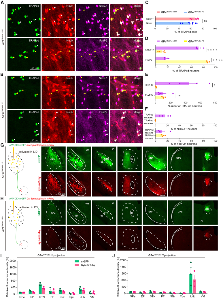
Activity history adds another layer to cell identity
Most captured GPe neurons express Nkx2.1, consistent with prototypic pallidal cells, whereas off-state-labeled neurons more often express FoxP2. The dyskinesia-related ensemble is only a small subset of all Nkx2.1 cells, adding activity history beyond molecular identity.
Tracing shows projections to STN and basal-ganglia output-related regions, with little return to striatum. Molecular features, output destinations and timing jointly characterize the ensemble, sharpening attention from the whole GPe to a particular subset.
Source: Shen C et al. · Cell Reports Medicine · CC BY-NC
Original figure and English caption ↗
A small ensemble can change the network’s motor outcome
This adult mouse study identifies ensembles through activity history and uses suppression and reactivation to distinguish accompaniment from behavioral contribution. It shows that pallidum contributes beyond relaying motor signals: a subset participates in abnormal movements and combines with striatal activity to shape severity.
Activity-dependent labeling mainly captures cells that become active, leaving suppressed populations outside this account. The result concerns part of the dyskinesia network, demonstrating that both anatomical location and the state in which cells are recruited matter for motor outcomes.