Why can one subthalamic nucleus produce opposing motor effects? Two populations meet different drug states
Increasing one population’s activity reduces dyskinesia, while another worsens some features. A male-mouse study distinguishes voluntary movement from abnormal involuntary actions.

An animal can move less yet show more abnormal involuntary actions, or have less dyskinesia without smoother voluntary movement. Do different populations within subthalamic nucleus contribute differently? A 2026 Science Advances study distinguishes populations projecting to entopeduncular and reticular tegmental nuclei and examines their activity and effects across levodopa states.
EP-defined activation reduces dyskinesia yet slows movement in other states; RtTg-defined activation worsens some dyskinetic features. Effects depend on the motor dimension and drug context.
Contents · 6 sections
Different outputs reveal internal differences
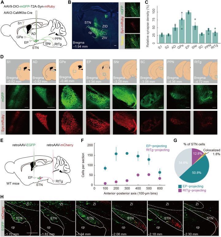
STN connects with several motor-related networks. The study compares cells projecting to entopeduncular nucleus (EP), within basal-ganglia output, and brainstem reticular tegmental nucleus (RtTg), associated with sensorimotor and cerebellar connections. Different outputs reveal potential functional differences within the region.
Overlap was limited, with EP-defined cells favoring anterior-dorsal STN and RtTg-defined cells posterior-ventral STN. Cells named by one output can also have other collaterals. These connection patterns let the team ask whether populations follow different trajectories within the same drug and behavioral state.
Source: Shen B et al. · Science Advances · CC BY
Original figure and English caption ↗
Movement and dyskinesia offer different windows
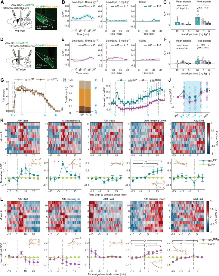
Dopamine-depleted male mice showed different motor patterns across levodopa states. Marked dyskinesia involved axial, limb and orolingual involuntary actions; other conditions increased movement with milder dyskinesia. Speed, turning and abnormal-action scores captured different aspects.
Overall STN mean and peak calcium signals did not differ significantly between the compared drug states, yet marked dyskinesia showed a U-shaped trajectory. Similar averages concealed different processes, encouraging the team to examine which populations stood out at each phase.
Two populations follow different trajectories
EP-defined activity followed a U-shaped pattern during marked dyskinesia, particularly around rising and falling phases. RtTg-defined activity mainly declined early and then recovered, with overall excitation in a milder state. Populations within one nucleus differed in timing and behavioral associations.
Speed, turning and time also shaped the relationships; accounting for them did not preserve every RtTg–dyskinesia correlation. Its signal may reflect broader action-related information. Functional interventions then asked how changing activity affected the animal’s movement.
Source: Shen B et al. · Science Advances · CC BY
Original figure and English caption ↗
Less dyskinesia need not mean faster movement
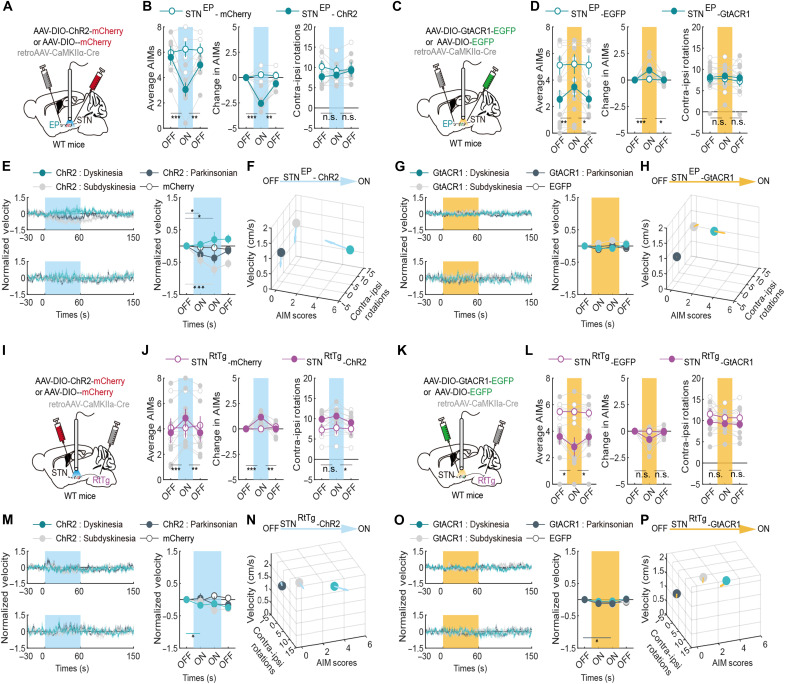
Increasing EP-defined activity lowered dyskinesia scores, which rebounded after stimulation; reducing activity raised them. Activation did not significantly change speed during marked dyskinesia but slowed movement and reduced walking in other states. Abnormal and voluntary movement diverged.
RtTg-defined activation worsened some dyskinetic features, particularly orolingual behavior. Reduced activity moderately lowered overall scores without changing every subscore. Speed and turning did not always follow scores, showing effects across different motor dimensions.
Source: Shen B et al. · Science Advances · CC BY
Original figure and English caption ↗
Different inputs and states converge
Input connectivity also differed. EP-defined cells had stronger associations with frontal, external-pallidal and parafascicular networks; RtTg-defined cells received more input from zona incerta, hypothalamus, bed nucleus of stria terminalis and periaqueductal gray. Distinct information sources accompanied distinct responses.
Without levodopa, altering either population did not induce score-defined dyskinesia, although walking and turning could change. Activity–behavior relationships depended on network state; the same intervention could produce different effects across backgrounds.
Motor effects emerge from population and state
The work separates a region’s motor effects into parallel populations, trajectories and movement dimensions. States with similar average activity can follow different phases, and lower dyskinesia can accompany slower movement. Population and state help explain seemingly opposing outcomes.
The findings concern adult male mice with unilateral injury and a drug model; population interventions also involve other collaterals. STN motor effects depend on connectivity, current state and ongoing movement. Its internal differences matter for understanding its overall contribution.