When an object moves, how does the brain map catch up? Sleep and septal–hippocampal memory timing
A mouse study finds that sleep loss changes how hippocampal maps update. Medial septal PV activity during encoding helps those maps adjust to a changing environment.
An object that occupied one corner yesterday now sits elsewhere. Recognizing that change requires remembering its old location while incorporating the new one into an active map. Sleep loss can disrupt this seemingly simple task. A 2026 Nature Communications study follows the medial septum (MS) and dorsal hippocampal CA1 (dCA1) in adult male mice. The problem extends beyond inactive neurons: how cells work together and when upstream signals arrive both matter for updating spatial memory.
Sleep deprivation weakens encoding-stage relationships between septal PV cells and hippocampal place cells. Enhancing septal–hippocampal activity during encoding improves field updating and object-location memory, unlike enhancement during rest or testing.
Contents · 6 sections
Still exploring, but less drawn to the new location
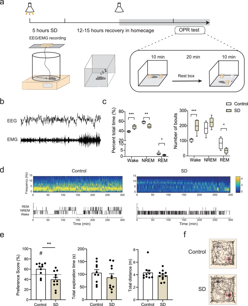
Control mice returning to a familiar arena preferentially explored the relocated object. This preference weakened after acute sleep deprivation. Sleep recordings showed reduced total and REM sleep and more wake bouts, while total exploration, distance and speed remained broadly unchanged. The memory difference emerged in animals that still moved and explored.
The task proceeded through habituation, location encoding, rest and testing. Whether an animal ultimately detected relocation did not identify the stage at which difficulty arose. Following activity across these phases revealed the handoff between remembering an old location and noticing a new one. Next-day stress and maze comparisons also showed no clear differences.
Source: Zheng Y et al. · Nature communications · CC BY-NC-ND
Original figure and English caption ↗
Septal cells form a population signal during object exploration
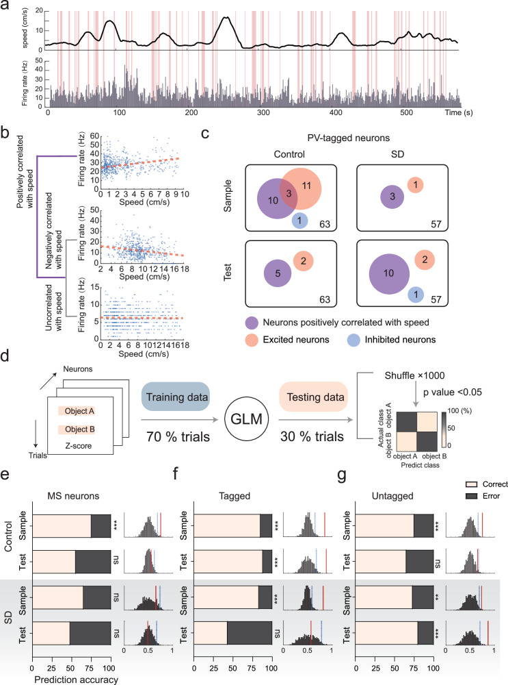
The medial septum is an important upstream contributor to hippocampal rhythms. Its PV cells were more active during encoding and testing, with a subset responding to object exploration. These cells were largely distinct from speed-related cells, and population activity did not closely track speed, shifting attention from how fast the animal moved to what it explored.
Individual PV cells weakly distinguished the two objects, but combining many cells made the difference clearer. This population discrimination declined at testing after sleep deprivation. Septal cells lacked the sharp spatial selectivity of hippocampal place cells, yet their collective activity carried information associated with objects and their locations.
Source: Zheng Y et al. · Nature communications · CC BY-NC-ND
Original figure and English caption ↗
The cells remain, but the map adjusts differently
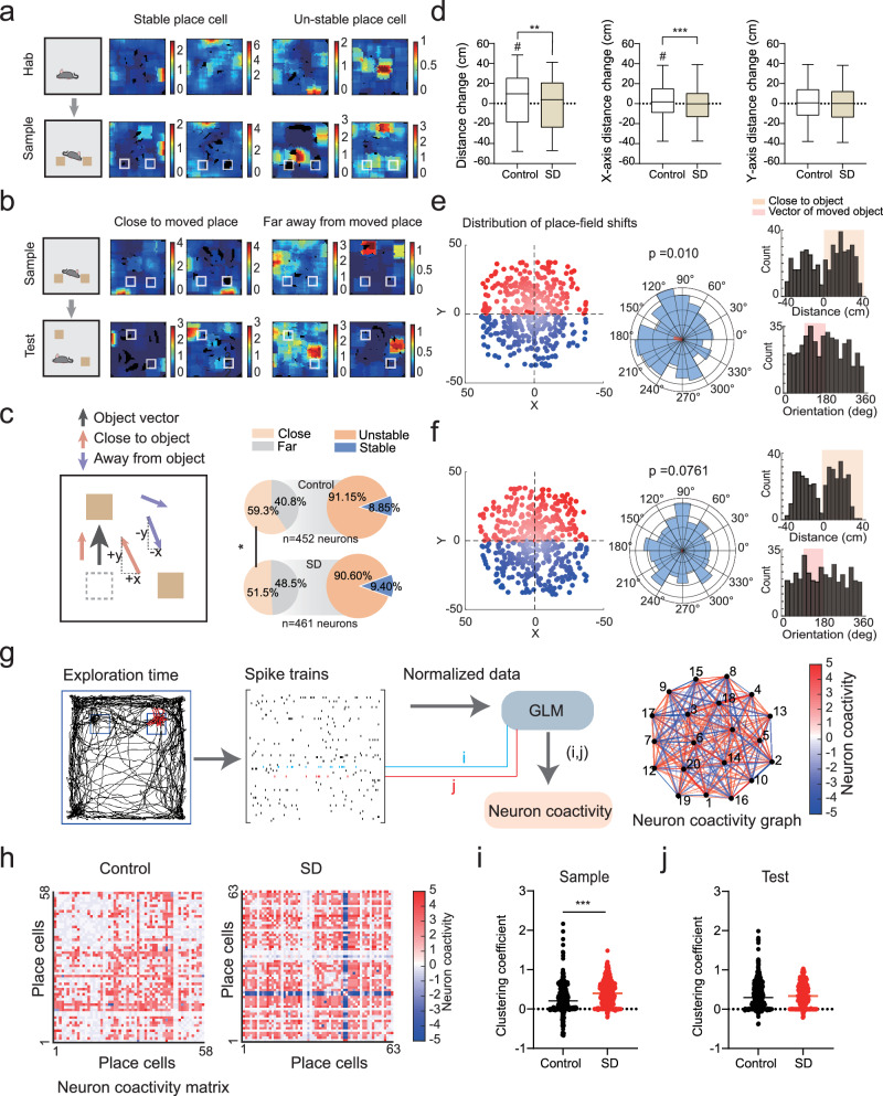
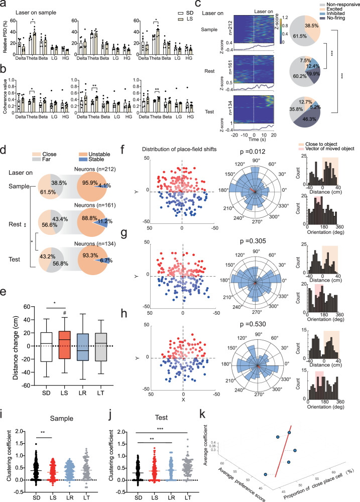
In controls, some dCA1 place fields shifted toward the relocated object, as though marking a newly important point on an existing map. Deprived mice lacked the same directional change. The proportions of place cells, remapping cells and unstable cells were comparable, placing the difference in how an existing map responded to environmental change.
Deprived mice also showed higher population coactivity during encoding. More cells acting together need not enrich information: excessive coupling among cells with distinct roles may reduce map flexibility, one interpretation proposed in the study. Familiarity and whether an object moved also affected cell relationships, making hippocampal organization task dependent.
Source: Zheng Y et al. · Nature communications · CC BY-NC-ND
Original figure and English caption ↗
Object-responsive septal cells relate to hippocampal maps
Tracking both regions revealed that object-responsive MS PV cells correlated with more dCA1 place cells during encoding. Different septal cells related to different place-cell subsets, resembling multiple parts within an upstream rhythm. Sleep deprivation weakened these relationships and their fine-grained coordination.
These relationships spanned different delays and involved indirect network processes. Existing work shows that septal GABA signals can regulate pyramidal activity through hippocampal inhibitory cells; theta rhythms and local networks may also contribute. The correspondence resembles coordination across several steps rather than one upstream cell directing one place cell.
Help arrives while locations are being encoded
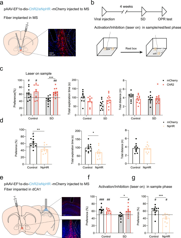
Could enhancing septal signals help deprived mice detect relocation again? Timing mattered. Enhancing MS PV activity during location encoding improved theta, septal–hippocampal coherence and field shifts, alongside changes in coactivity structure. Mice again showed a preference for the relocated object.
Enhancement during rest or testing did not restore preference. Suppressing PV activity at several phases could impair performance, and broad suppression also reduced test exploration. Septal–hippocampal cooperation spans the task, but encoding offers a particularly effective rescue window after sleep disruption; enhancement at the final test arrives too late for this benefit.
Source: Zheng Y et al. · Nature communications · CC BY-NC-ND
Original figure and English caption ↗
An output linking rhythms with location memory
Focusing on MS PV terminals in dCA1 showed that enhancing this output during encoding also improved post-deprivation location memory, whereas suppression impaired performance. This links septal cells, hippocampal maps and exploration through a specific pathway. Other septal outputs, including those toward entorhinal cortex, remain part of distributed spatial processing.
This study of adult male mice concerns a specific location-memory task. Its implication is concrete: updating memory needs both appropriate cell relationships and appropriate timing. After sleep loss, place cells remain and animals continue exploring. What changes is how the map incorporates new cues and whether septal signals organize its timing during encoding.
Source: Zheng Y et al. · Nature communications · CC BY-NC-ND
Original figure and English caption ↗