Two signals from hippocampal CA3: how does CCK support spatial learning?
Beyond fast excitation, CA3 cells provide a peptide signal. A mouse study follows the CA3–CA1 pathway to connect CCK release, spatial exploration and synaptic potentiation.
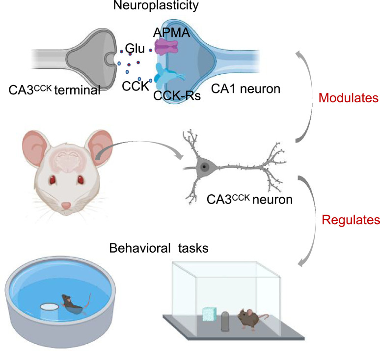
Finding a platform or noticing a moved object engages hippocampal information processing. What role do peptide signals play alongside fast transmission? Researchers examined excitatory CCK cells in dorsal CA3, following their CA1 output, evoked peptide release, learning-related activity and contribution to performance and potentiation.
Suppressing CA3 CCK output or reducing Cck expression weakens spatial performance and long-term potentiation, revealing peptide modulation within an excitatory pathway.
Contents · 6 sections
A familiar CCK marker points to another population
Cholecystokinin is a peptide signal and a marker of neuronal populations. In the hippocampus it is often associated with inhibitory interneurons, yet shared peptide expression does not imply the same fast transmitter. This study examines excitatory CCK cells in dorsal CA3 and their contribution to CA3-to-CA1 transmission and plasticity.
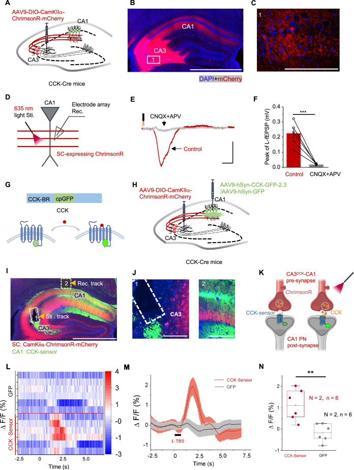
Reporter co-labeling identified CCK-positive cells associated with excitatory markers in CA3, including cells projecting to CA1. A peptide often associated with inhibitory interneurons thus led to an excitatory output and a more focused question about spatial learning.
One pathway carries excitation and a peptide signal
Stimulating labeled CA3 CCK projections evoked CA1 field excitatory potentials largely blocked by glutamate receptor antagonists. With a CCK sensor in CA1, theta-burst optical stimulation in CA3 produced a transient fluorescence increase absent from GFP controls. The pathway thus supports excitatory transmission accompanied by peptide release.
The transient peptide signal added a modulatory dimension: CA3 cells provide glutamatergic output and can deliver CCK to CA1. Because CA1 receives multiple CCK sources, the next step was to follow this identified population during behavior.
Source: Huang F et al. · eLife · CC BY
Original figure and English caption ↗
Activity changes from early learning to familiar navigation
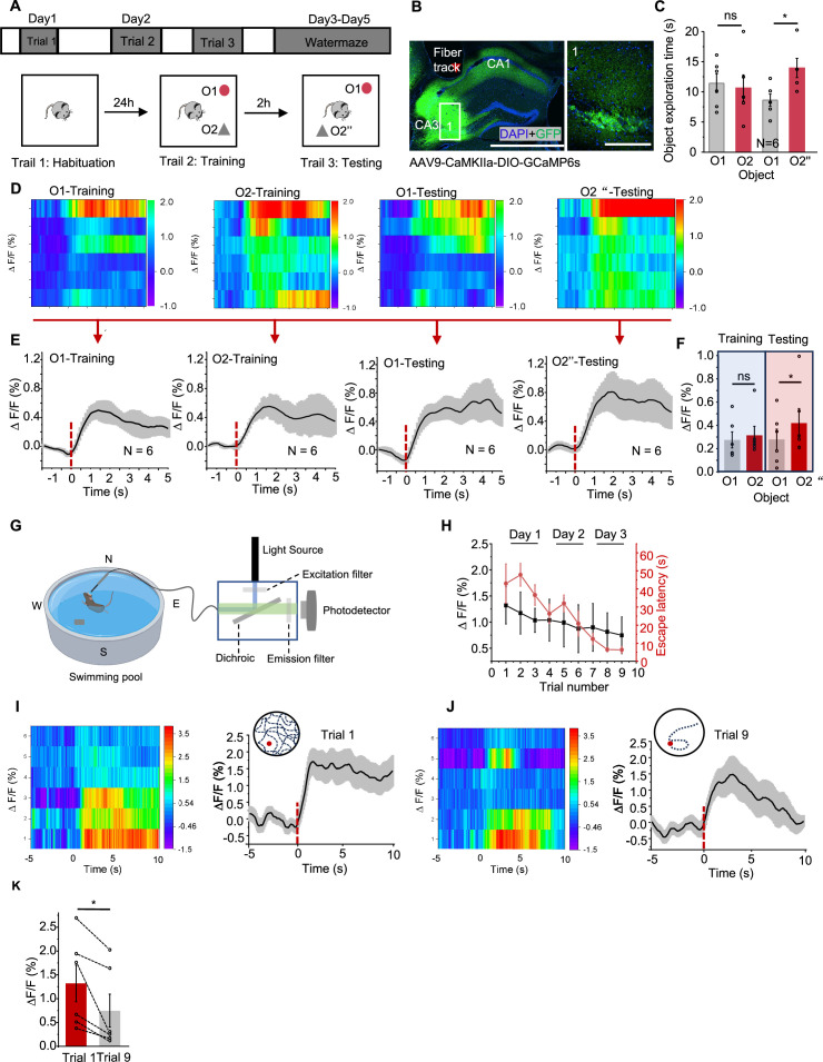
In object-location testing, exploration increased calcium signals in excitatory CA3 CCK neurons, with a larger response to the novel location during testing. In the water maze, initial hidden-platform learning produced stronger, more sustained activity than a later well-trained trial. Activity therefore varied with learning stage and spatial exploration.
Initial exploration and familiar platform finding differ in novelty, routes and task demands. The population recordings linked CA3 CCK activity to learning context and prompted a functional test: would weakening the output change location discrimination and platform learning?
Source: Huang F et al. · eLife · CC BY
Original figure and English caption ↗
Weaker output changes two spatial tasks
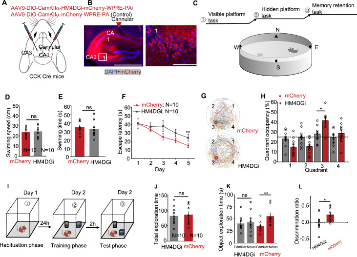
An inhibitory tool was expressed in the relevant CA3 cells, with local CA1 drug delivery targeting their terminals. Hidden-platform performance and target-quadrant preference deteriorated, and object-location novelty preference was lost relative to controls. The convergent tasks support a contribution of this output to normal spatial performance.
Swimming speed, visible-platform performance and total exploration remained comparable. Slice inhibition also lowered baseline field responses and reduced theta-burst potentiation. Potentiation persisted at a smaller magnitude, suggesting that the output supports full plasticity.
Source: Huang F et al. · eLife · CC BY
Original figure and English caption ↗
A further question: does CCK itself contribute?
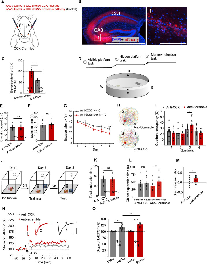
Because output inhibition affects multiple signals, the team next reduced Cck expression in these CA3 cells. Verified mRNA reduction accompanied poorer hidden-platform learning and location discrimination, while swimming, visible-platform and exploration controls remained unchanged. This extends the interpretation from cell output to the peptide itself.
After Cck reduction, ordinary electrically evoked field responses remained comparable while potentiation declined. This separation highlighted a modulatory role in activity-dependent strengthening. Parallel spatial and synaptic effects made the peptide a concrete entry point into learning mechanisms.
Source: Huang F et al. · eLife · CC BY
Original figure and English caption ↗
Peptide modulation accompanies fast transmission in spatial learning
The study links CA3 cells, CA1 output, evoked CCK release, spatial exploration and potentiation across scales. CCK can accompany excitatory output as well as occur in inhibitory cells, adding a layer of modulation associated with normal spatial performance and full potentiation.
Behavioral evidence comes from male mice. Natural CA1 peptide sources, receptor action and finer cell specificity remain open, with whole-cell and intersectional approaches offering ways forward. Their coordination with fast transmission across learning stages is a promising next question.