Two mammillary populations divide place and object recognition
In mice, excitatory PV and Drd2 mammillary neurons connect with different hippocampal inputs and thalamic outputs, supporting distinct pathways during place and object recognition testing.
A familiar object moved elsewhere and a new object appearing in the same place are different changes. Mice investigate novelty, but how does the brain distinguish where from what? A study published in Advanced Science in 2025 examines the mammillary body, often viewed as a hippocampal–thalamic relay. PV- and Drd2-marked medial mammillary neurons differ in molecular features, firing, connectivity and recognition behavior, forming functionally differentiated memory-related routes.
Two excitatory medial mammillary populations connect with dorsal or ventral subiculum and different thalamic regions. PV-related interventions primarily alter place recognition and Drd2-related interventions object recognition, with test-phase activity modulation affecting novelty preference.
Contents · 7 sections
A memory node contains internal divisions
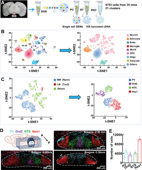
Single-cell transcriptional analysis resolves multiple mammillary cell clusters, with medial neurons divided into PV, Drd2, neurotensin and Nos1 types. Tissue mapping reveals distinct spatial distributions rather than uniform mixing.
The study focuses on PV and Drd2 populations, both with excitatory glutamatergic characteristics. PV in this region differs from the inhibitory populations commonly associated with the marker elsewhere. A marker can participate in different connectivity and functional contexts across regions.
Source: Li L et al. · Advanced science (Weinheim, Baden-Wurttemberg, Germany) · CC BY
Original figure and English caption ↗
Spontaneous firing and rebound bursts distinguish the populations
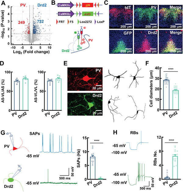
PV neurons are larger and fire spontaneously at rest. Drd2 neurons are smaller and largely quiet at rest, but readily generate rebound bursts after hyperpolarization. These excitatory populations differ in how their firing responds to input and state changes.
The populations also differ in transcriptional features, including multiple genes associated with electrical activity. Their classification therefore extends beyond labeling and position to how they generate signals, potentially contributing to memory-related specialization.
Source: Li L et al. · Advanced science (Weinheim, Baden-Wurttemberg, Germany) · CC BY
Original figure and English caption ↗
Gene interventions change selected firing patterns
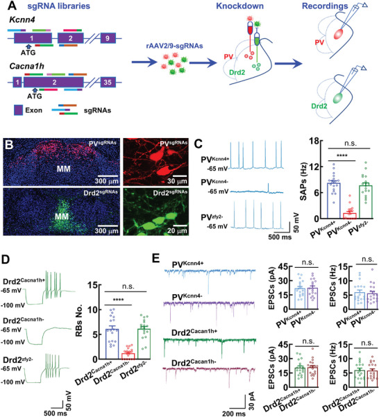
Lowering Kcnn4 expression in PV cells markedly reduces spontaneous firing; lowering Cacna1h in Drd2 cells reduces rebound bursts. Each intervention affects a characteristic firing pattern without completely eliminating activity.
Measured miniature excitatory-current amplitude and frequency do not significantly change. The altered firing patterns are thus distinguished from these synaptic-input readouts, providing different activity perturbations for subsequent place- and object-memory comparisons.
Source: Li L et al. · Advanced science (Weinheim, Baden-Wurttemberg, Germany) · CC BY
Original figure and English caption ↗
Place and object changes are affected differently
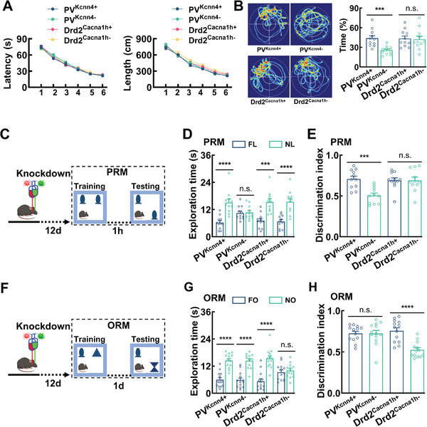
During place recognition, a familiar object moves to a new location. The PV-related intervention reduces preference for that location, without a corresponding Drd2 effect. During object recognition, a new object appears in the same place; the Drd2-related intervention reduces novel-object preference, without a significant corresponding PV effect.
The discrimination index is time investigating the novel target divided by total investigation of both targets. A value near one half indicates little preference; a higher value indicates more investigation of novelty. The ratio places location and object changes on a common behavioral scale.
The place and object tests use one-hour and one-day delays, respectively. With shorter delays, affected groups still show novelty preference. Water-maze training paths and latencies remain similar, while PV-related probe performance is weaker. These contrasts emphasize delayed recognition rather than a general loss of exploration ability.
Source: Li L et al. · Advanced science (Weinheim, Baden-Wurttemberg, Germany) · CC BY
Original figure and English caption ↗
The populations connect with different upstream and downstream regions
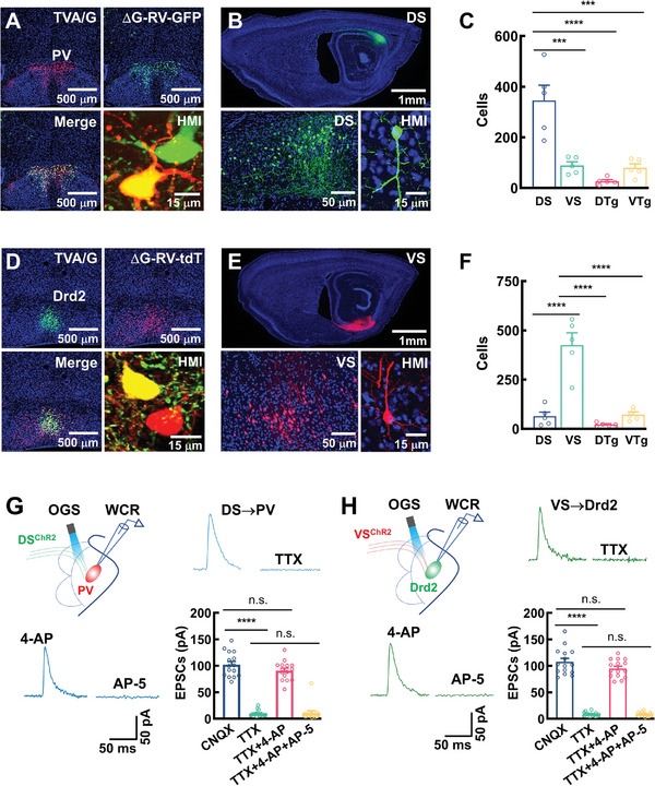
Tracing and slice recordings identify predominantly dorsal-subicular input to PV cells and ventral-subicular input to Drd2 cells. Terminal-evoked excitatory currents and blockade-and-rescue experiments support direct connectivity, linking input origins with the population distinction.
Downstream tracing identifies preferential PV output to anteroventral thalamus and Drd2 output to anteromedial thalamus. These corresponding routes connect hippocampal output, mammillary cell types and thalamic targets, resolving a traditional relay into more specific circuitry.
Source: Li L et al. · Advanced science (Weinheim, Baden-Wurttemberg, Germany) · CC BY
Original figure and English caption ↗
Recruiting the relay population improves measured preference
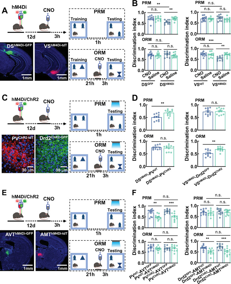
Suppressing the two subicular terminal pathways impairs place or object recognition, respectively. Activating the corresponding mammillary population during testing improves preference, whereas training-phase activation does not alter measured performance. Timing clarifies the routes’ contribution during recognition tests.
Suppressing the corresponding thalamic region counteracts improvement from mammillary activation. Input perturbation, relay activation and downstream inhibition converge on an interregional relationship, extending beyond an association between performance and a cellular marker.
Source: Li L et al. · Advanced science (Weinheim, Baden-Wurttemberg, Germany) · CC BY
Original figure and English caption ↗
Novel locations and objects recruit different calcium responses
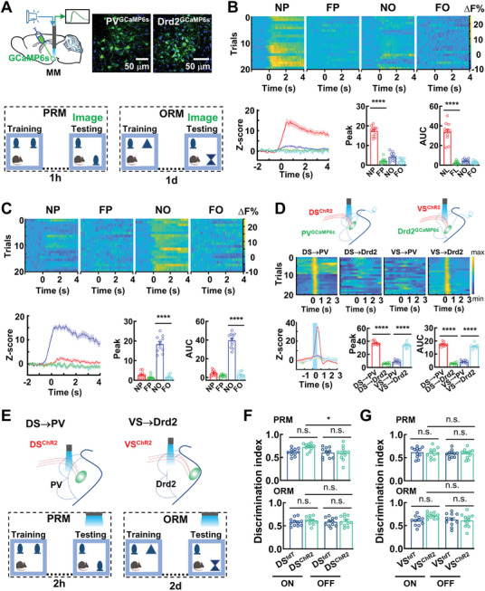
During testing, PV-population calcium responds more strongly to a novel location, and Drd2 calcium to a novel object. Familiar locations and objects lack the same prominent changes, matching the distinctions from behavioral interventions.
With longer test delays, activating the corresponding subicular input increases preference for a novel location or object, without the same change in no-light conditions. This male mouse study reveals differentiated mammillary signaling: distinct cells connect different inputs and outputs and separately influence place and object recognition.
Source: Li L et al. · Advanced science (Weinheim, Baden-Wurttemberg, Germany) · CC BY
Original figure and English caption ↗