Plasticity at a hippocampal output: CCK and two converging inputs
In male mice, coactivating CCK-labeled entorhinal input and CA1 input strengthens CA1-to-distal-subiculum responses. Local suppression of the associated input in subiculum also reduces object-location discrimination.
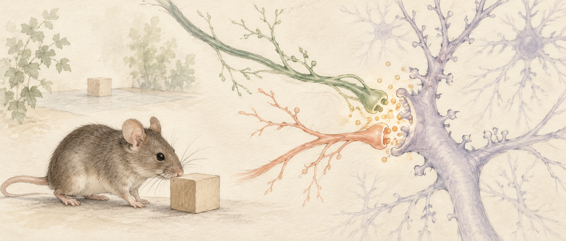
When an object moves, a mouse spends more time investigating it. How does the brain retain the original location and respond to change? A study in Communications Biology focuses on the subiculum, a hippocampal output region. Coactivating cholecystokinin, or CCK-labeled entorhinal input with CA1 input strengthens the latter’s synaptic response. A molecule familiar from digestion and satiety also contributes to neural plasticity.
CCK and coactivity of two inputs support long-term potentiation in the CA1-to-distal-subiculum pathway. Associated entorhinal cells respond during object exploration, while local input suppression reduces location-discrimination measures.
Contents · 7 sections
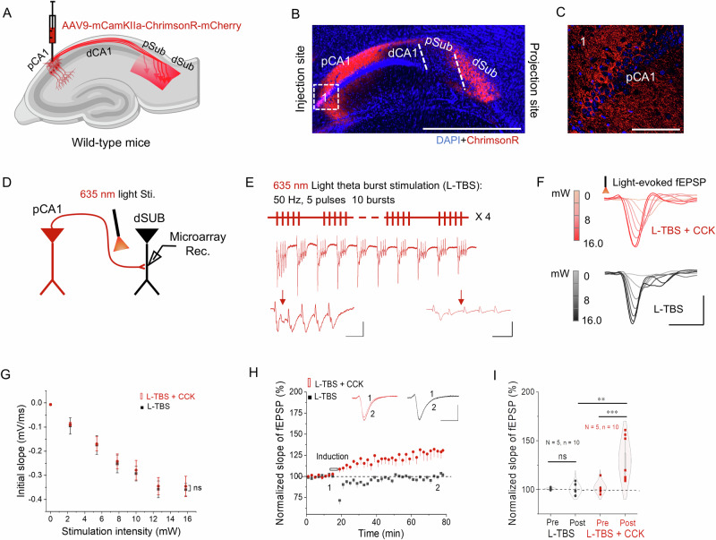
A single input fails to potentiate until CCK is added
The subiculum receives CA1 and entorhinal input and is an important route for hippocampal information to other regions. Synaptic plasticity concerns whether the same input evokes a stronger response after a period of activity. The study compares field responses from CA1 input in distal subiculum slices.
Recruiting CA1 input alone does not produce significant long-term potentiation under the tested conditions. With the added CCK fragment CCK-4, subsequent responses to the input become stronger. The comparison suggests that neuropeptide modulation influences whether activity leaves a lasting change in the connection.
Source: Huang F et al. · Communications biology · CC BY-NC-ND
Original figure and English caption ↗
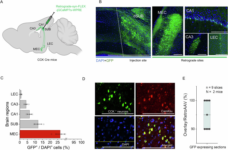
Entorhinal cortex supplies CCK-labeled input
Tracing CCK-labeled sources to distal subiculum identifies projecting cells in several regions, with higher labeled-cell density in medial entorhinal cortex, or MEC. Most of these cells carry an excitatory marker, and their subicular input has a clear glutamatergic component. CCK labeling here accompanies excitatory long-range connectivity.
Recruiting the MEC-derived input alone likewise does not produce significant potentiation under the tested slice conditions. Origin and CCK labeling alone do not describe the eventual change, so the team turns to the state created when the two inputs meet.
Source: Huang F et al. · Communications biology · CC BY-NC-ND
Original figure and English caption ↗
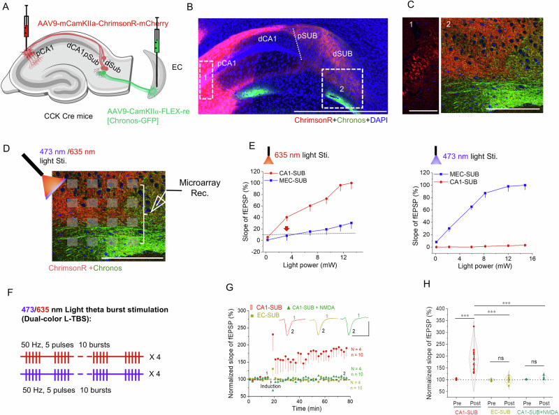
CA1 input potentiates while both inputs do not strengthen together
Coactivation of CA1 and CCK-labeled MEC input markedly strengthens CA1-to-subiculum field responses, persisting through subsequent recording. MEC-to-subiculum responses themselves show no significant potentiation. Activity in one input population affects the efficacy of another.
This cross-input change is termed heterosynaptic long-term potentiation. NMDA-receptor blockade prevents significant potentiation during coactivation, linking coordination between inputs with a receptor-dependent plasticity process. Here, long-term describes the persistence measured during experimental recording.
Source: Huang F et al. · Communications biology · CC BY-NC-ND
Original figure and English caption ↗
Subicular CCK signals rise after input recruitment
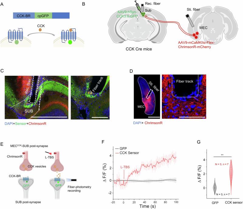
The team next asks whether subiculum shows a CCK signal during MEC-input activity. Sensor recordings in anesthetized mice show higher CCK-associated fluorescence after recruitment, without the same change in a conventional fluorescent control. This supports input-associated local neuropeptide release.
The elevation persists for a period, while sensor dynamics also depend on receptor and reporter response kinetics. It reveals a neuropeptide-modulation layer which, alongside rapid glutamatergic transmission, supplies molecular context for connection changes after coactivation.
Source: Huang F et al. · Communications biology · CC BY-NC-ND
Original figure and English caption ↗
Baseline responses are similar while potentiation is impaired
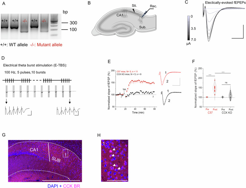
A separate comparison uses CCK-deficient mice. Baseline electrically evoked field responses are similar to controls, but induced potentiation is impaired and not significant in the deficient group. Transmitting a signal and strengthening after activity are different functions of a connection.
Tissue labeling also identifies CCK-B receptors in subiculum. An exogenous CCK fragment, local release signals, and gene-loss results connect CCK with plasticity from different angles, extending attention from input strength to the modulatory conditions provided by a neuropeptide.
Source: Huang F et al. · Communications biology · CC BY-NC-ND
Original figure and English caption ↗
Exploration evokes responses and local suppression affects discrimination
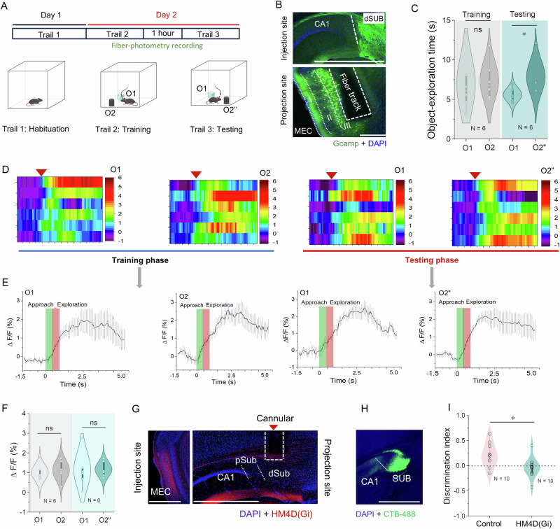
MEC CCK-labeled cells projecting to subiculum show transient calcium responses during exploration of both objects. After an object moves, mice explore the new location more, but response peaks for the two objects do not differ significantly. The population participates in exploration without simply mirroring location preference.
Reducing the associated MEC input locally in subiculum lowers discrimination between new and familiar object locations. This complements the synaptic observations: input activity affects location-task performance, while CCK modulation relates to connection plasticity. The study uses adult male mice.
Source: Huang F et al. · Communications biology · CC BY-NC-ND
Original figure and English caption ↗
Plasticity emerges through coordination between inputs
The study views subiculum as a site of convergence and modulation. CA1 input, entorhinal input, and neuropeptide context jointly influence how a connection changes after activity, while the same recruitment conditions can have different effects on the two inputs.
The observations place connection strengthening in a context of converging information and neuropeptide modulation. Exploration and plasticity experiments separately reveal behavioral involvement and connection changes, adding a neuropeptide perspective to regulation of hippocampal output.