Turning light into a local hippocampal signal: polydopamine particles and mouse spatial memory
From light absorption to cellular responses and brain rhythms, a study follows how photothermal conversion relates to spatial-task performance across different states.
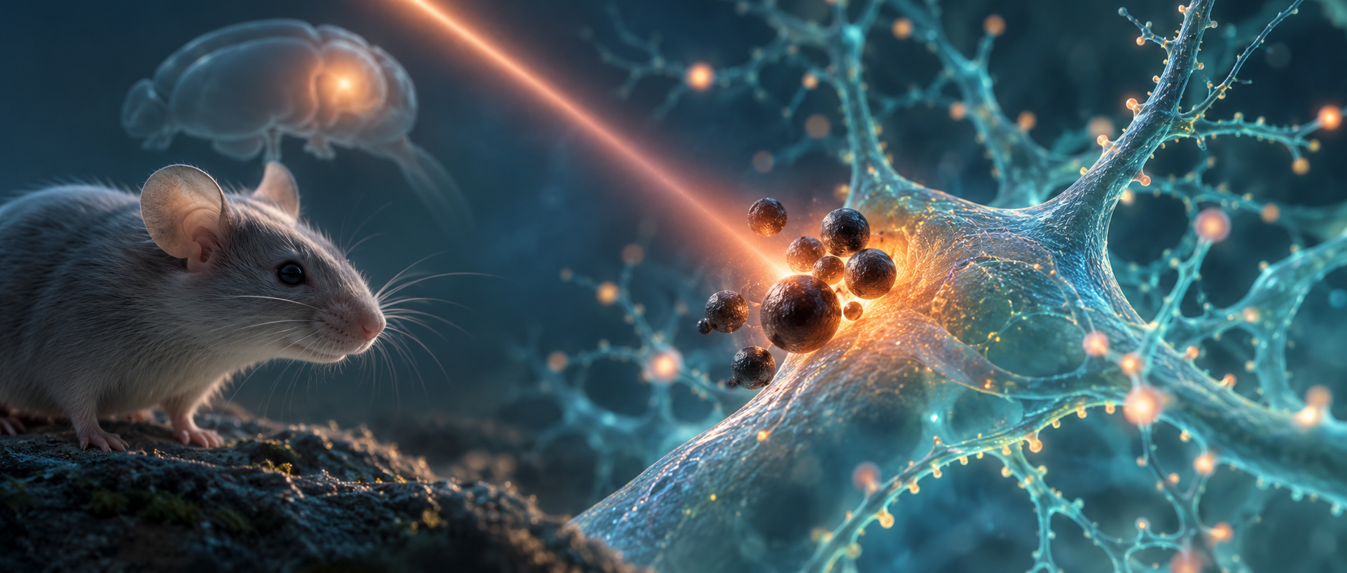
Light is absorbed and scattered as it enters tissue. Reaching deeper cells requires sufficient energy and a way to turn it into a usable local signal. A 2026 Advanced Science study assigns that conversion to polydopamine particles: in a local region of the mouse dentate gyrus, particles absorb near-infrared light and generate a thermal response. The team followed calcium activity, firing and rhythms to examine how this physical conversion related to spatial-memory tasks.
Particles combined with near-infrared light alter local activity and improve Barnes-maze performance, without significant Y-maze improvement. TRPV1-related effects and model-dependent gamma changes connect the material with cellular and network responses.
Contents · 7 sections
The material absorbs light and converts energy
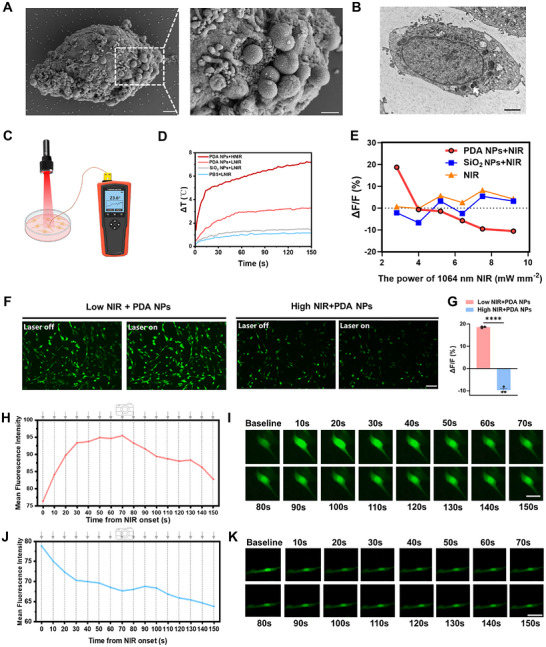
Polydopamine takes its name from dopamine polymerization, but its role here is photothermal. Near-infrared light produces a stronger thermal response where it meets local particles. Material location provides the regional focus and allows comparisons with particle-free conditions.
Under the tested conditions, cell-viability, hemolysis and tissue observations showed no clear injury signal, and particles remained locally for an extended period. The next question concerned function: how would a thermal change become a cellular response?
Changing energy changes the cellular response
PC12 cells provided the first answer. Lower-power light combined with particles increased calcium fluorescence, whereas higher power decreased it. Light alone and similarly sized silica particles did not produce the same response. The outcome depended jointly on material and energy.
The cellular experiments also distinguished interpretations. No clear dopamine-monomer release occurred at lower power, favoring photothermal effects over dopamine release from the material. PC12 offered a model for separating physical and cellular effects before examining a brain network.
Source: Zhou YB et al. · Advanced science (Weinheim, Baden-Wurttemberg, Germany) · CC BY
Original figure and English caption ↗
A temperature-sensitive channel connects heat with cellular signals
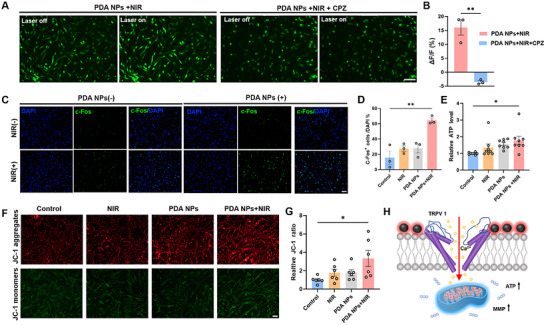
Temperature-sensitive TRPV1 became a connecting point. Blocking it reduced the calcium response to combined particles and light. Stimulation also accompanied changes in activity-related c-Fos, mitochondrial membrane potential and ATP, bringing heat, channel activity and energy state into one interpretation.
The authors propose that local warming influences calcium entry through TRPV1 and couples with mitochondrial state. The model explains how a material’s physical properties might influence cells and focuses attention on the links producing the response.
Source: Zhou YB et al. · Advanced science (Weinheim, Baden-Wurttemberg, Germany) · CC BY
Original figure and English caption ↗
Calcium and firing change together in dentate gyrus
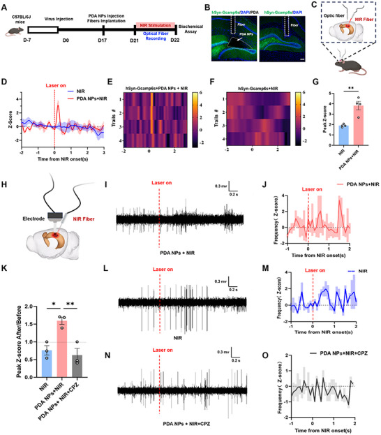
In mouse dentate gyrus, the team examined calcium signals and local electrical activity. Both increased under the combined condition, without equivalent clear changes under near-infrared light alone. TRPV1 blockade also weakened the firing effect, connecting the cellular-channel clue with brain activity.
Activity-related labeling concentrated near the particle-containing region, linking the site of light–material interaction with response distribution. The effect involves a local network of different cells, shaped by the actual distributions of particles and temperature.
Source: Zhou YB et al. · Advanced science (Weinheim, Baden-Wurttemberg, Germany) · CC BY
Original figure and English caption ↗
Spatial-task improvement varies across memory measures
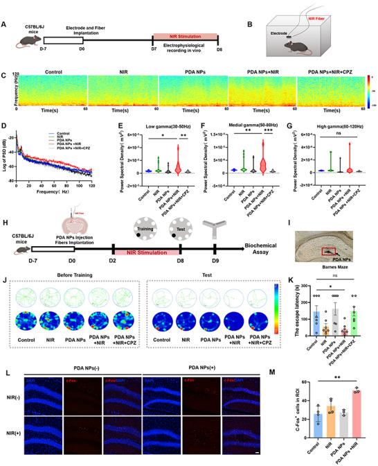
Normal mice showed increased low- and middle-gamma activity without significant high-gamma change. The combined group found the target hole faster in the Barnes maze; TRPV1 blockade removed this advantage and reduced related rhythm changes. Local activity, rhythms and task performance converged.
Another spatial measure did not improve in parallel: Y-maze spontaneous alternation showed no significant group difference. Although the light-only group had a shorter average Barnes time, it lacked the same clear statistical improvement. Outcomes varied with task and condition.
Source: Zhou YB et al. · Advanced science (Weinheim, Baden-Wurttemberg, Germany) · CC BY
Original figure and English caption ↗
The same strategy evokes different rhythms across states
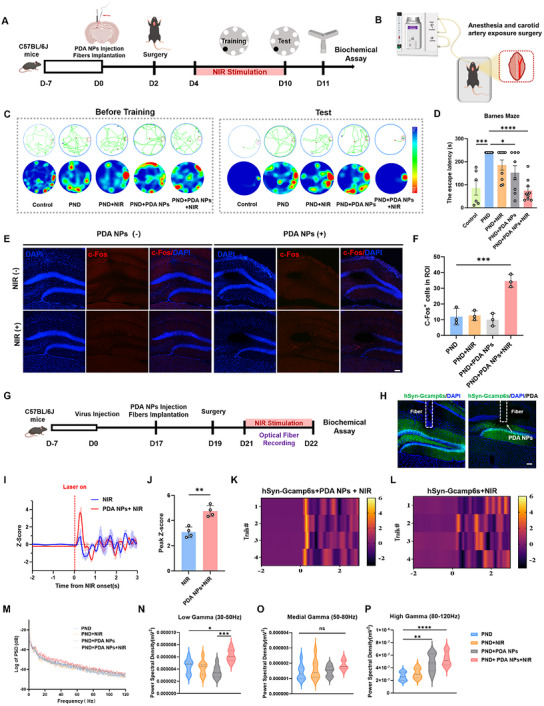
In the perioperative neurocognitive-disorder model, the combined condition also shortened Barnes latency and increased dentate calcium activity and c-Fos. The gamma pattern differed: low and high bands increased, while the middle band did not change significantly. The same strategy met a different network state.
Y-maze performance again showed no significant improvement, and particles alone also changed some Barnes and high-gamma measures. The authors relate the latter to possible antioxidant or anti-inflammatory material effects, adding another potential contribution alongside combined illumination.
Source: Zhou YB et al. · Advanced science (Weinheim, Baden-Wurttemberg, Germany) · CC BY
Original figure and English caption ↗
Physical conversion meets neural networks
The study connects a physical process with observable cellular and network changes: absorbed light produces a local thermal response, alongside channel-related effects and altered neural activity. Spatial-task results show that animal state and task also shape the network’s response.
The evidence comes from PC12 cells and adult male mice, with particles placed locally within the brain. It demonstrates a research strategy under specific conditions: considering where light is converted, how cells respond and what task the animal performs explains more than activity enhancement alone.