The brain’s brakes can strengthen: CCK cells modulate auditory cortex and sound–threat association
A mouse study finds that cholecystokinin-expressing inhibitory cells can strengthen their own and another cell class’s inhibitory inputs. Activating them after training reduces sound-evoked freezing the next day.
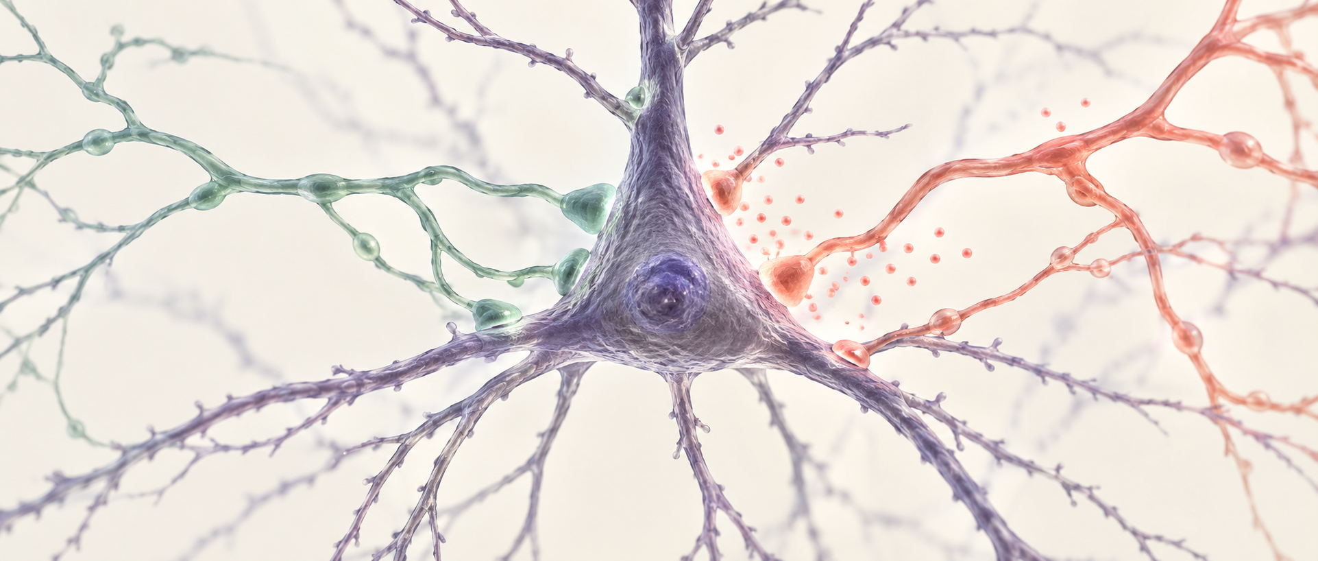
Learning is often associated with stronger connections among neurons, but what strengthens need not be excitation; it can be inhibition. A study published on August 25, 2025 in Scientific Reports identifies inhibitory long-term potentiation in mouse auditory cortex. Cholecystokinin-expressing GABAergic cells can strengthen inhibition onto pyramidal cells and help another inhibitory class strengthen its output. Linking this cellular account with sound–shock association, the study finds that activating CCK cells after training reduces sound-evoked freezing the next day.
Under the tested conditions, CCK inhibitory cells more readily induce sustained inhibitory potentiation than PV or SST cells. Exogenous CCK enables potentiation of the latter inputs, while effects on sound-associated behavior differ by cell class.
Contents · 6 sections
Inhibitory strength can retain a sustained change
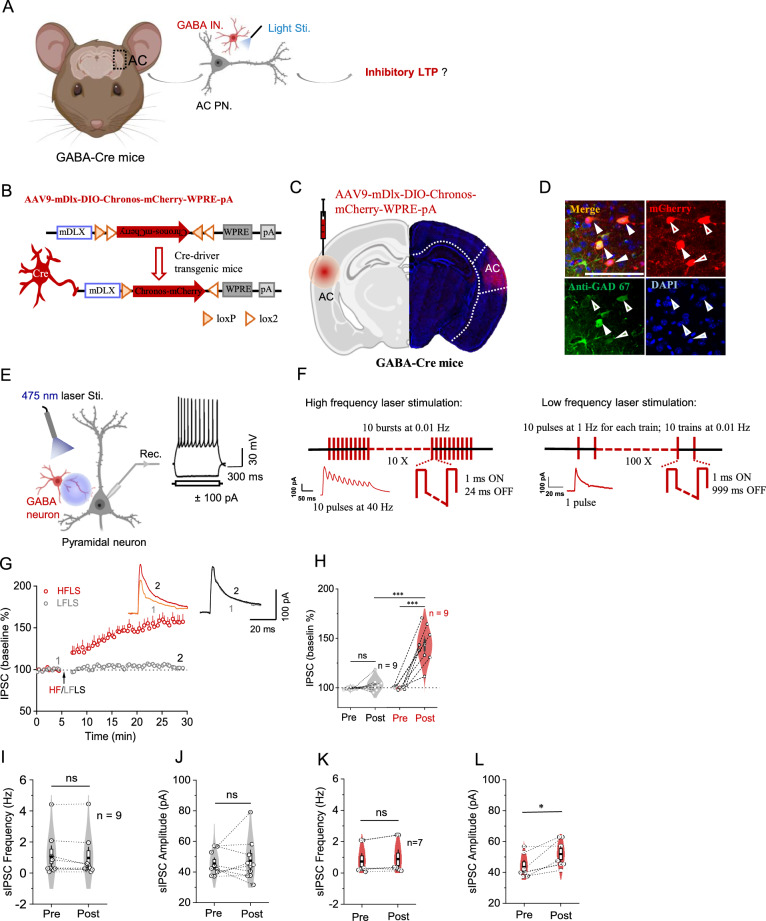
Auditory-cortical pyramidal cells receive excitatory input and inhibition from GABAergic cells. Enhancing a broad GABAergic population with a high-frequency pattern produces a sustained increase in evoked inhibitory postsynaptic currents on pyramidal cells; a lower-frequency pattern lacks this change. Inhibition can strengthen and remain elevated during subsequent recording.
Spontaneous inhibitory events offer another view. After high-frequency activation of the broad GABAergic population, event amplitude increases while frequency remains broadly unchanged. The change concerns individual event strength rather than merely more frequent events, motivating the question of which inhibitory class contributes.
Source: Zhang G et al. · Scientific reports · CC BY-NC-ND
Original figure and English caption ↗
The same activation pattern yields a different result for CCK
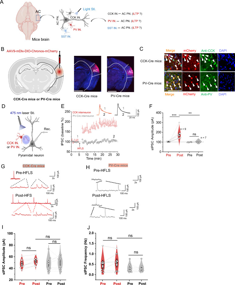
The study compares CCK-, PV- and SST-expressing inhibitory cells. Under the same high-frequency activation conditions, CCK-to-pyramidal evoked inhibition potentiates, whereas PV and SST inputs retain broadly similar strength. CCK-associated potentiation appears in superficial and deep layers, extending beyond one cortical layer.
Notably, selective CCK-cell activation does not noticeably increase the amplitude or frequency of all spontaneous inhibitory events recorded on pyramidal cells. Selectively potentiated inputs and spontaneous events pooling multiple sources can differ. Cortical inhibition is not a single control knob; cell classes and observation scales matter.
Source: Zhang G et al. · Scientific reports · CC BY-NC-ND
Original figure and English caption ↗
With CCK, PV and SST inputs can also potentiate
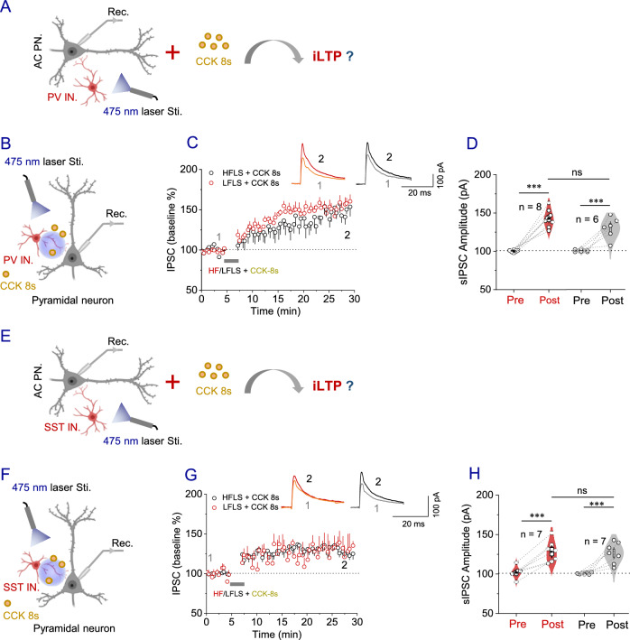
CCK is not just a cell label; it is a neuropeptide. In the presence of exogenous CCK, PV and SST inhibitory inputs that previously lacked clear potentiation now strengthen. Both higher- and lower-frequency activation paired with CCK produce sustained changes of comparable magnitude.
Supplementary experiments find that CCK alone can also induce inhibitory potentiation. The peptide thus changes the modulatory conditions around synapses, allowing previously unpotentiated inputs to become plastic. How CCK is released during natural activity and which specific receptors mediate its effects remain key links to ordinary auditory processing.
Source: Zhang G et al. · Scientific reports · CC BY-NC-ND
Original figure and English caption ↗
CCK cells can support stronger PV inhibition
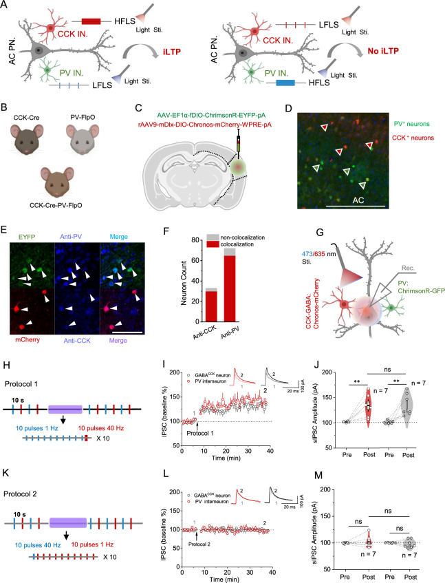
If CCK changes the plasticity of other inputs, can CCK-cell activity help nearby PV input? Separately controlling the classes shows that high-frequency CCK activity paired with low-frequency PV activity potentiates both inhibitory currents onto pyramidal cells. This change across inputs is termed heterosynaptic plasticity.
Reversing the activity allocation—high frequency in PV and low frequency in CCK—fails to produce equivalent potentiation in either input. The effect depends on how activity is distributed across classes, rather than any simultaneous recruitment. CCK-cell influence extends beyond its own contacts to another inhibitory input converging on pyramidal cells.
Source: Zhang G et al. · Scientific reports · CC BY-NC-ND
Original figure and English caption ↗
Post-training CCK activation reduces next-day freezing
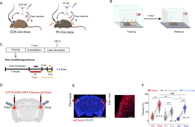
How does cellular inhibitory potentiation relate to behavior? Mice learn a sound–shock association, receive auditory-cortical CCK-cell activation during early post-training consolidation, and hear the sound in a different context the following day. They freeze less than controls but more than before training, indicating an attenuated associative response.
Equivalent PV-cell activation yields no clear difference in next-day freezing relative to controls. Cell class matters in both synaptic and behavioral experiments. Timing matters too: the experiment modulates an established association during early consolidation rather than directly facilitating movement while the sound is played.
Source: Zhang G et al. · Scientific reports · CC BY-NC-ND
Original figure and English caption ↗
Inhibitory cells do more than suppress activity immediately
The study extends inhibition beyond an immediate brake to a network process capable of sustained adjustment and interactions. CCK cells potentiate their own input and help PV input change, while the peptide enables plasticity in additional inhibitory classes. Learning-related changes can therefore involve both excitatory and inhibitory sides.
These findings come from mouse auditory-cortical slices and a sound-association task. Whether potentiation is expressed presynaptically or postsynaptically, and how it contributes to the behavioral change, require more direct tests. The results identify a possibility: local inhibitory networks can participate in shaping association strength by changing their own plasticity.