How does a magnetic field influence oxytocin? PIEZO2 connects the molecular clues in mice
Magnetic-nanoparticle-assisted stimulation produces opposing effects at different frequencies, linking a mechanosensitive channel, oxytocin release and social exploration in a mouse model.
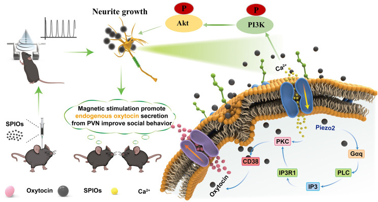
In a three-chamber task, mice can investigate an unfamiliar conspecific or a nonsocial area. This choice offers a window into social behavior. A study published on July 8, 2025 in the Journal of Nanobiotechnology finds that magnetic-nanoparticle-assisted stimulation of the hypothalamic paraventricular nucleus (PVN) changes oxytocin release, increases social exploration and reduces excessive grooming in mice exposed to valproic acid prenatally. The mechanosensitive ion channel PIEZO2 emerges as a key molecular contributor. Magnetic stimulation does not invariably promote these effects: lower-frequency stimulation shifts oxytocin and behavior in the opposite direction.
Reducing PVN PIEZO2 prevents higher-frequency magnetic stimulation from restoring oxytocin release, cellular calcium responses and associated behavioral improvements, supporting a role for this mechanosensitive channel.
Contents · 7 sections
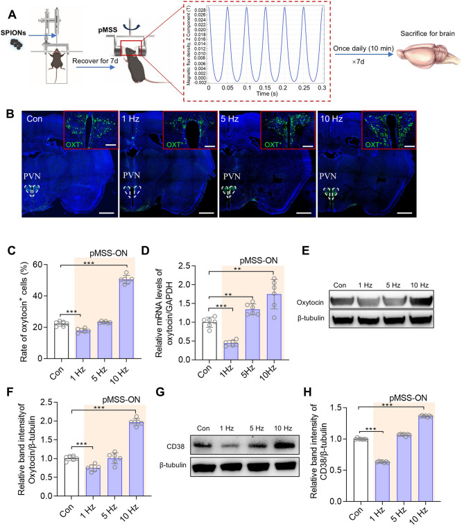
Oxytocin changes depend on stimulation conditions
The PVN is an important source of oxytocin in the brain. The study introduces magnetic nanoparticles into this region before using a changing magnetic field to modulate local activity. Its question is specific: can the combination alter cells’ capacity to release their own oxytocin?
The answer depends on frequency. Lower-frequency conditions reduce oxytocin-related expression, whereas higher-frequency conditions increase it, together with the secretion-associated protein CD38. An external physical signal thus produces bidirectional biological effects, with the direction closely tied to stimulation conditions.
Source: Liu S et al. · Journal of nanobiotechnology · CC BY-NC-ND
Original figure and English caption ↗
Behavioral changes persist through follow-up
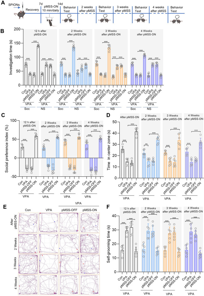
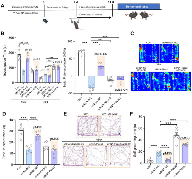
Model mice initially investigate conspecifics less, spend less time in the open-field center and groom for longer. Higher-frequency stimulation increases social investigation and preference, increases center exploration and reduces excessive grooming. These are behavioral changes across distinct tasks; approaching a conspecific does not itself establish complex social understanding.
The changes appear at the first assessment and persist during subsequent weeks through a one-month follow-up. The opposite result is also striking: lower-frequency stimulation in control mice reduces social investigation and center time while increasing grooming. These opposing behavioral directions parallel the oxytocin-related changes.
Source: Liu S et al. · Journal of nanobiotechnology · CC BY-NC-ND
Original figure and English caption ↗
Mechanosensitive PIEZO2 comes into focus
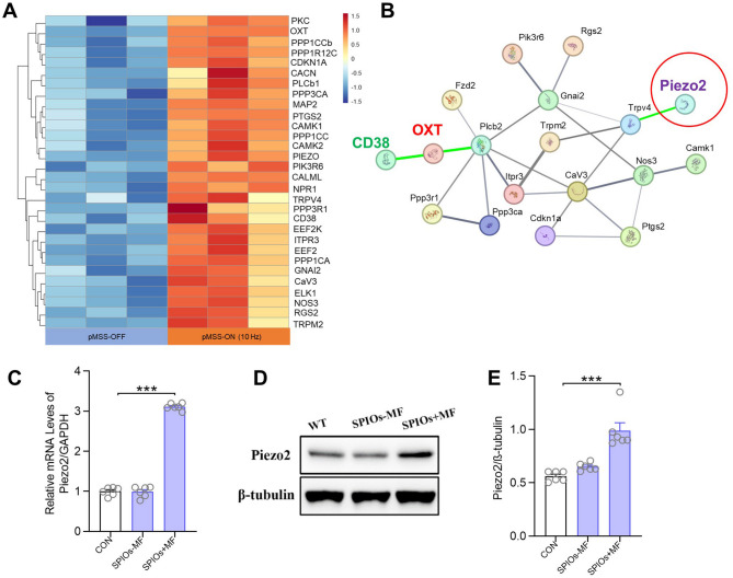
A question separates the magnetic field from neural secretion: how does a cell translate a physical change into an internal signal? Gene analysis identifies several candidate ion channels, and subsequent validation focuses attention on PIEZO2. Higher-frequency stimulation increases its expression, whereas several other candidate channels show no corresponding significant change.
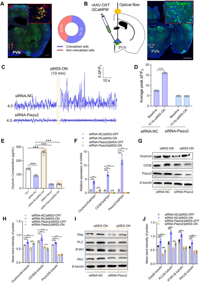
PIEZO2 senses mechanical influences and belongs to a class of channels that translate force into cellular signals. Imaging shows overlapping localization with a subset of PVN oxytocin cells. This establishes a testable connection: a mechanosensitive channel may participate in neuroendocrine changes triggered by nanoparticle-assisted magnetic stimulation.
Source: Liu S et al. · Journal of nanobiotechnology · CC BY-NC-ND
Original figure and English caption ↗
Reducing the channel prevents calcium and secretion increases
Higher-frequency stimulation increases calcium signals in oxytocin cells and oxytocin release in the PVN. Reducing PIEZO2 in this region lowers baseline cellular calcium responses and oxytocin release; subsequent magnetic stimulation fails to reinstate the original increases.
This result moves PIEZO2 from a candidate that changes alongside stimulation to an important contributor to its effects. It also shows that the channel supports ongoing cell function: reducing it affects the cell’s state before stimulation as well as its response to the external signal.
Source: Liu S et al. · Journal of nanobiotechnology · CC BY-NC-ND
Original figure and English caption ↗
Neurites in cultured cells also respond
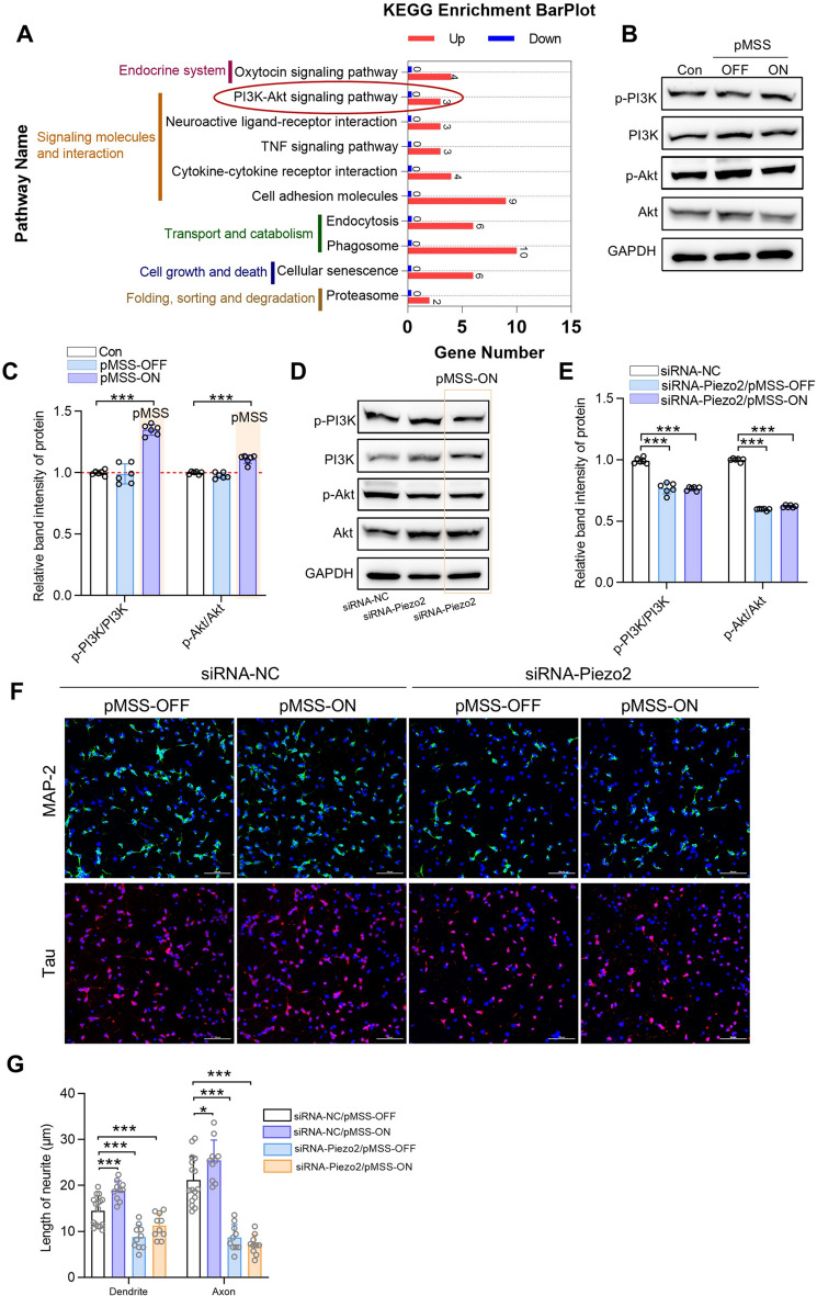
The changes extend beyond secretion. Higher-frequency stimulation increases phosphorylation signals in the PVN PI3K–Akt pathway, which is associated with cellular growth and plasticity. Reducing PIEZO2 weakens these signals, and stimulation fails to restore them.
In cultured hypothalamic neurons from model mice, stimulation is accompanied by longer dendrites and axons. PIEZO2 reduction inhibits neurite growth, which stimulation does not restore. These cell experiments reveal a structural response, although they do not directly establish whether neurite elongation sustains the behavioral changes over subsequent weeks.
Source: Liu S et al. · Journal of nanobiotechnology · CC BY-NC-ND
Original figure and English caption ↗
The molecular link converges with social exploration
Finally, the study returns to the mice’s choices. Reducing PVN PIEZO2 further decreases social investigation and increases grooming in model mice; higher-frequency stimulation loses its earlier capacity to improve these behaviors. Changes in cellular signals, oxytocin release and behavior thus converge under the same molecular perturbation.
This does not make one hormone responsible for all social behavior. The study also observes correlations between oxytocin release and some behavioral measures. How oxytocin, the channel and growth pathways each contribute remains a question to resolve individually.
Source: Liu S et al. · Journal of nanobiotechnology · CC BY-NC-ND
Original figure and English caption ↗
From magnetic particles to intracellular responses
In a prenatal-exposure model with male and female mice, the study connects nanoparticle-assisted magnetic stimulation to a mechanosensitive channel. Frequency changes the direction, PIEZO2 influences the response, and oxytocin and growth-related signals participate. The route from an external physical change into the cell gains a more concrete molecular explanation.
Social exploration and repetitive behavior in this model cover only part of a complex neurodevelopmental phenotype. Responses in models with different etiologies, together with the long-term effects of particles in the brain, will determine how far this mechanism extends.