Sound as a second hit: an auditory route amplifies vulnerability in a Parkinsonian mouse model
Loud sound unmasks motor deficits in model mice with initially normal performance. An auditory-to-nigral connection and a dopamine vesicle-loading protein link environmental input to neuronal vulnerability.
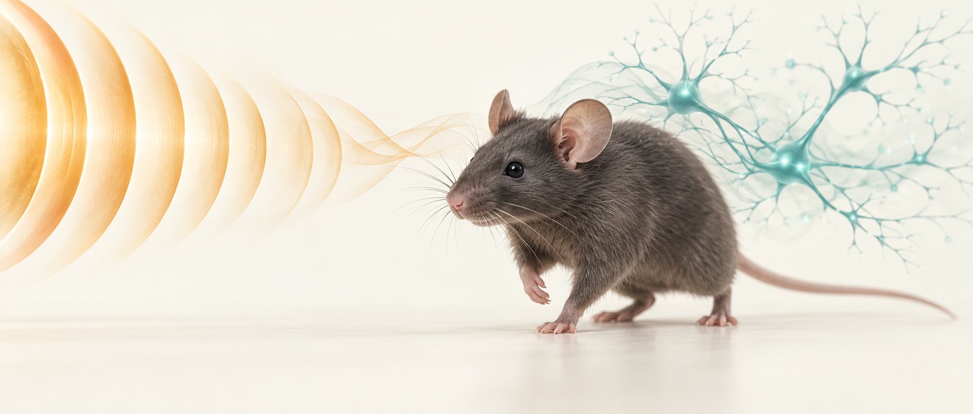
Why might hearing a sound affect a step? Auditory input does not remain within the hearing system. A study published in PLOS Biology on November 4, 2025 finds that loud sound can reveal vulnerability in mice with mild dopaminergic injury but no overt motor deficits. Changes after one exposure recover quickly; repeated exposure leaves deficits detectable a week later. Following an inferior-colliculus-to-substantia-nigra connection, the study identifies a molecular link involving the vesicular monoamine transporter VMAT2.
Inferior colliculus directly connects to nigral dopaminergic neurons. Modulating these auditory-input recipients mimics or attenuates loud-sound-associated motor changes; increasing their VMAT2 improves movement and attenuates the reduction in nigral TH-positive cells.
Contents · 7 sections
One loud exposure temporarily disrupts motor performance
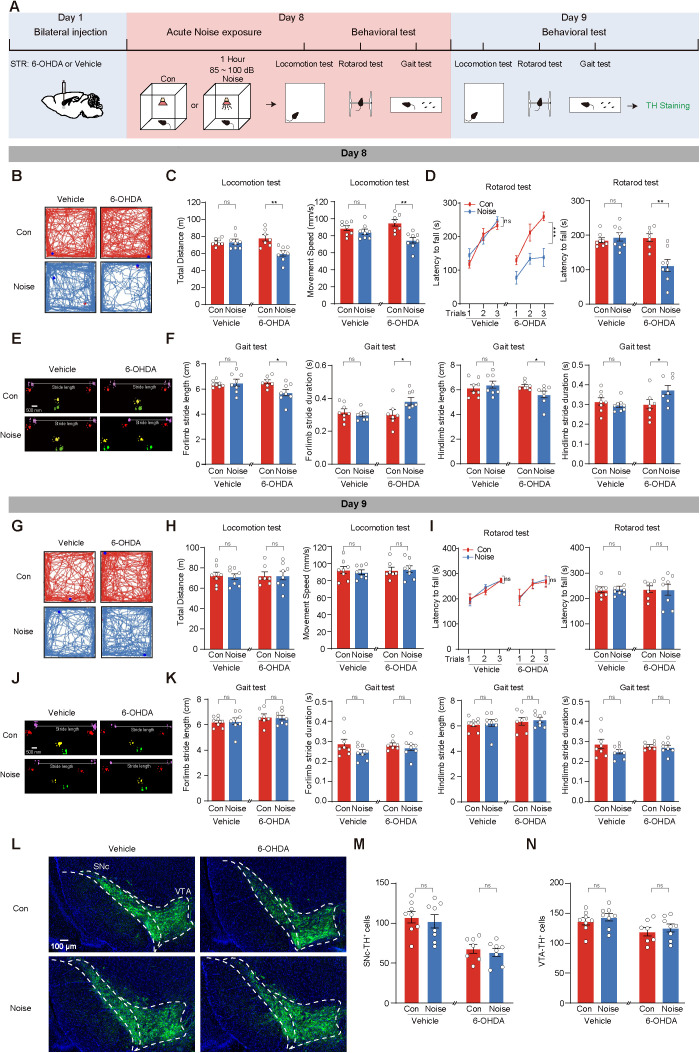
The study starts at an informative point: low-dose 6-OHDA partially injures nigral dopaminergic cells while motor tests remain comparable to controls. This places the question before overt deficits, asking whether external input can disrupt performance. After loud white-noise exposure, these mice travel less, move more slowly, fall sooner from a rotarod and show altered gait.
By the next day, motor measures are again comparable to controls, and nigral TH-positive cell counts are not further reduced relative to injury alone. Uninjured mice retain broadly unchanged motor performance under the same sound conditions. A single loud exposure therefore first manifests as functional disruption in an already vulnerable system rather than immediate additional detectable cell loss.
Source: Cui C et al. · PLoS biology · CC BY
Original figure and English caption ↗
Motor difficulties remain a week later
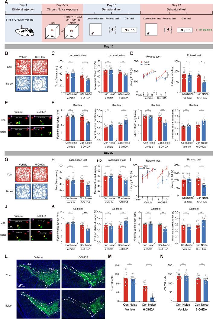
Repeated loud exposure changes the picture. After consecutive days of exposure, model mice still move less and more slowly the following day, with poorer rotarod and gait performance; these changes remain at the one-week test. Nigral TH-positive cells are further reduced, whereas counts in neighboring ventral tegmental area are comparable, indicating regional differences.
Control mice do not develop equivalent motor deficits, and other Parkinsonian animal models provide supporting findings. Replacing white noise with music at similarly high sound levels still changes movement in model mice. Everyday labels such as music or noise are therefore less informative here than the experimental combination of loud sound and existing neural injury.
Source: Cui C et al. · PLoS biology · CC BY
Original figure and English caption ↗
Inferior colliculus sends input to substantia nigra
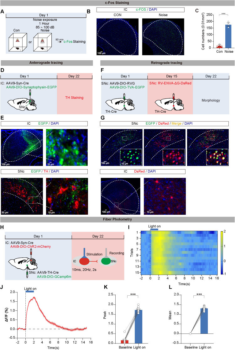
Inferior colliculus is a major midbrain auditory relay. Loud sound increases its activity marker and enhances firing in recorded putative nigral dopaminergic cells. Anatomical tracing shows a direct connection from collicular cells to nigral dopaminergic neurons, allowing auditory input to enter a system closely linked to movement.
Functional measurements show what the connection can do: enhancing inferior-collicular activity elevates calcium signals and firing in nigral dopaminergic cells. Auditory and motor pathways intersect here. To understand why stronger input accompanies poorer movement, the study next asks whether activity in these recipient cells determines the injured model’s response to sound.
Source: Cui C et al. · PLoS biology · CC BY
Original figure and English caption ↗
Modulating auditory-input recipients changes the second hit
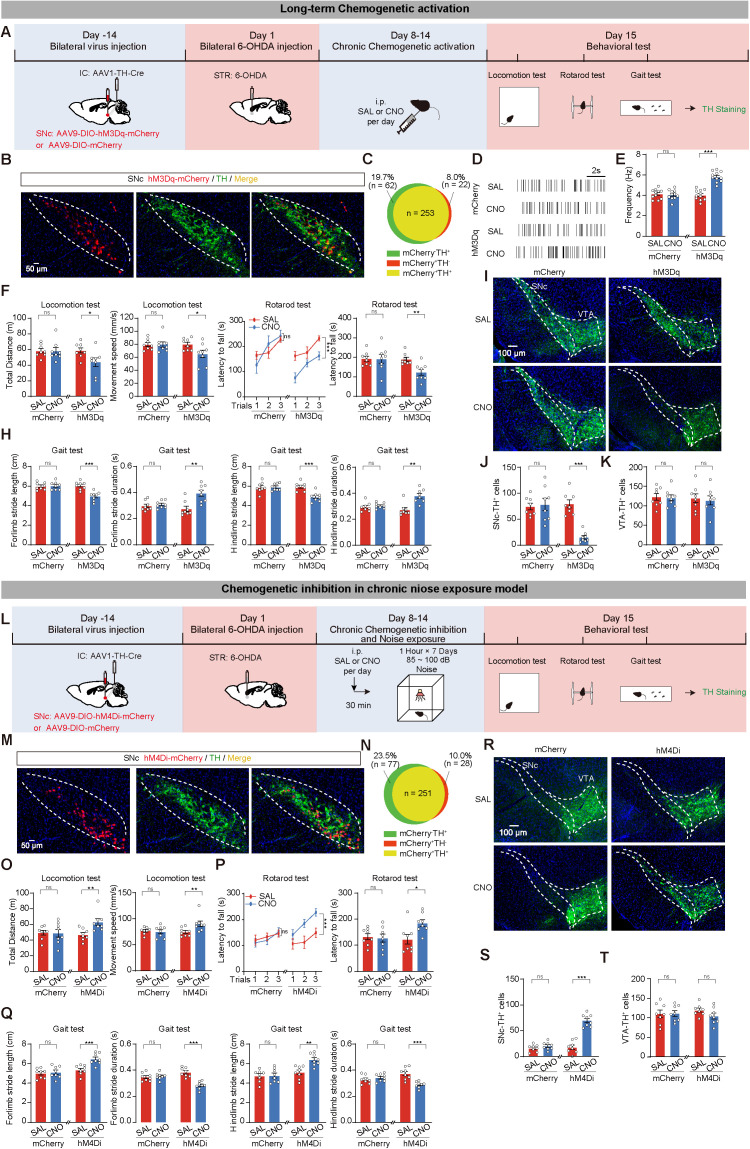
Briefly enhancing collicular-input-recipient nigral dopaminergic cells produces motor difficulties in injured mice even without loud sound, without additional TH-positive cell reduction. Conversely, suppressing these cells during acute sound exposure improves locomotion, rotarod and gait performance. The functional changes are bidirectionally modifiable.
Sustained enhancement accompanies poorer motor performance and reduced nigral TH-positive cells. Suppressing these cells during repeated sound exposure both improves movement and attenuates the reduction in cell counts. Short-term functional disruption and later cellular changes converge on cells recruited through the same connection.
Source: Cui C et al. · PLoS biology · CC BY
Original figure and English caption ↗
VMAT2 connects activity changes to intracellular handling
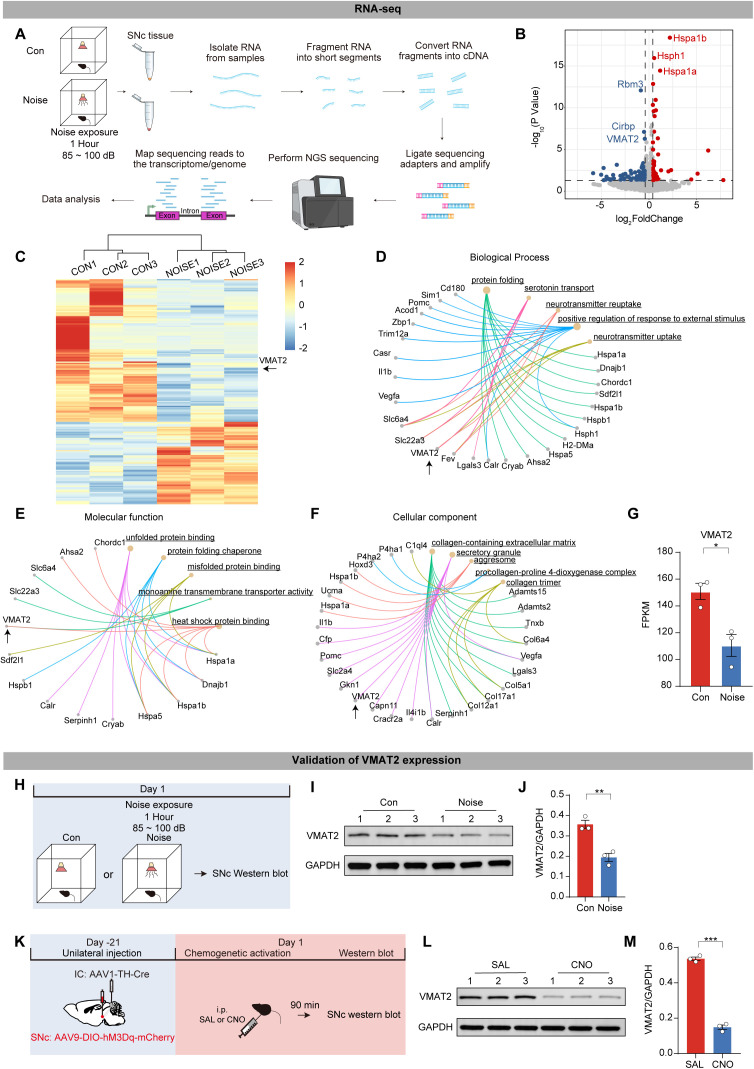
Neurons must not only produce dopamine but load it into vesicles for signaling. VMAT2 performs this loading task. Gene-expression analysis of nigral tissue after sound exposure identifies changes involving protein folding, vesicle secretion and transmitter transport, with reduced VMAT2 standing out as one lead.
Protein measurements likewise find reduced VMAT2, and enhancing collicular-input-recipient nigral cells produces this change even without sound exposure. This connects pathway activity to intracellular dopamine handling. Earlier work on VMAT2 also links vesicular loading to transmitter storage and neuronal resilience under injury.
Source: Cui C et al. · PLoS biology · CC BY
Original figure and English caption ↗
Increasing VMAT2 improves resilience to loud sound
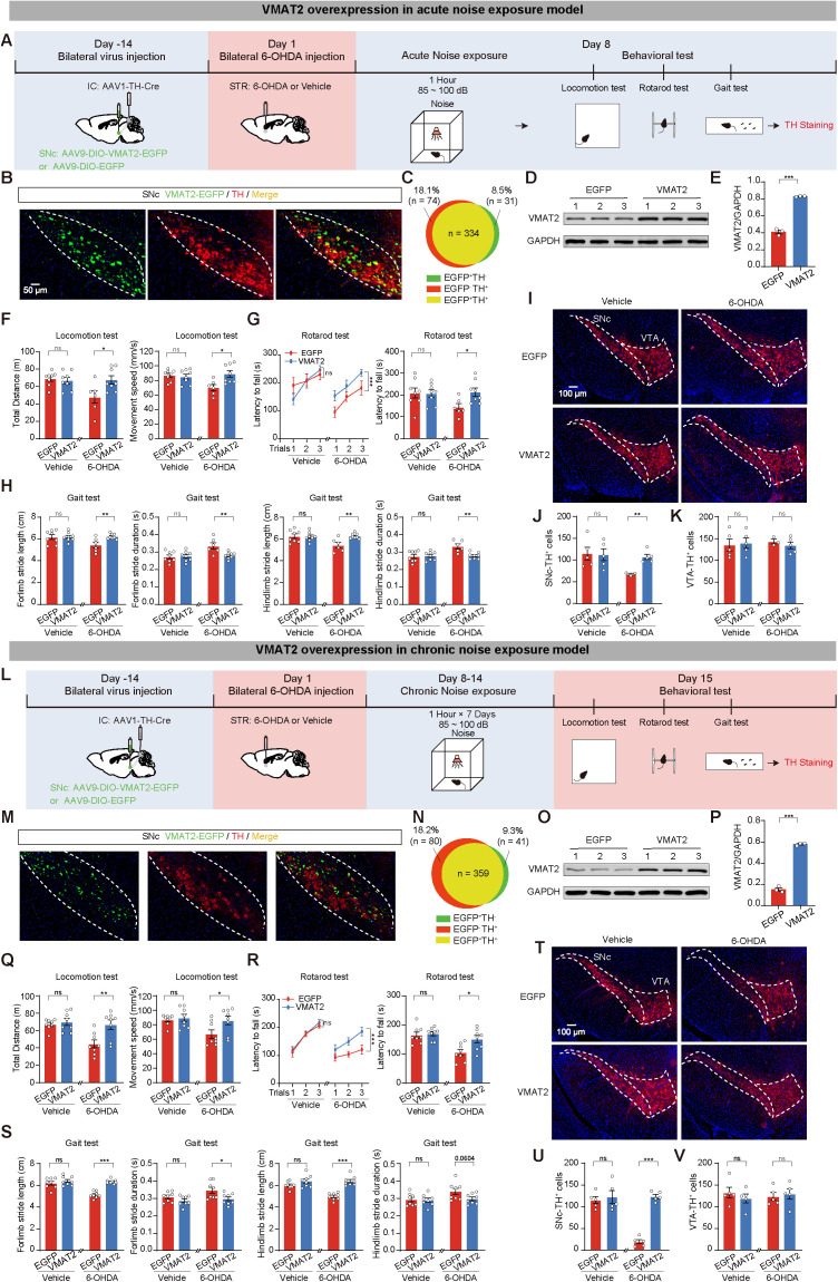
If VMAT2 merely accompanied the changes, increasing it might not improve outcomes. The study therefore raises VMAT2 in nigral dopaminergic cells receiving collicular input. After one loud exposure, injured mice show improved distance traveled, speed, rotarod and gait performance. Repeated-exposure conditions likewise show better movement and more nigral TH-positive cells.
This restoration experiment links the molecular lead to behavior. External sound can alter cellular activity through neural connections, while transmitter loading relates to the system’s capacity to withstand a second challenge. Sound can also recruit stress-related processes beyond this auditory route, and other molecules identified in nigral tissue may contribute.
Source: Cui C et al. · PLoS biology · CC BY
Original figure and English caption ↗
Environmental input can shift balance before overt deficits
The study places motor-system vulnerability in the context of sensory input. A system that maintains performance in routine tests can be transiently disrupted by additional loud sound, with repeated challenges leaving more persistent changes. An auditory relay may help determine how existing injury becomes manifest.
These observations come from adult male mice and specific injury models, with sound levels above ordinary quiet surroundings. They reveal an interaction among environmental input, neural activity and intracellular transmitter handling: existing injury does not act alone, as the system continually responds to external signals.